. 2010 May 20;6(5):e1000911.
doi: 10.1371/journal.ppat.1000911.

Neisseria meningitidis Opc invasin binds to the sulphated tyrosines of activated vitronectin to attach to and invade human brain endothelial cells

Affiliations

Neisseria meningitidis Opc invasin binds to the sulphated tyrosines of activated vitronectin to attach to and invade human brain endothelial cells

Claudia Sa E Cunha et al. PLoS Pathog. .

Abstract

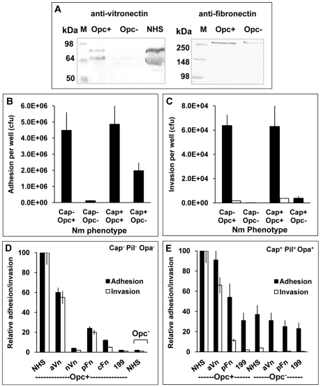
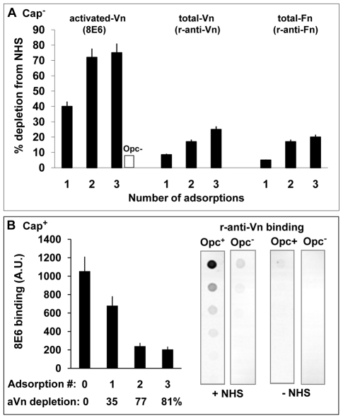
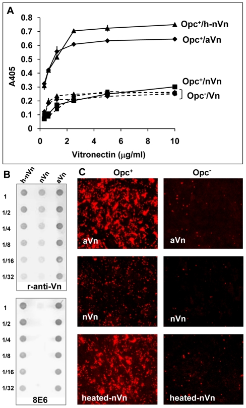
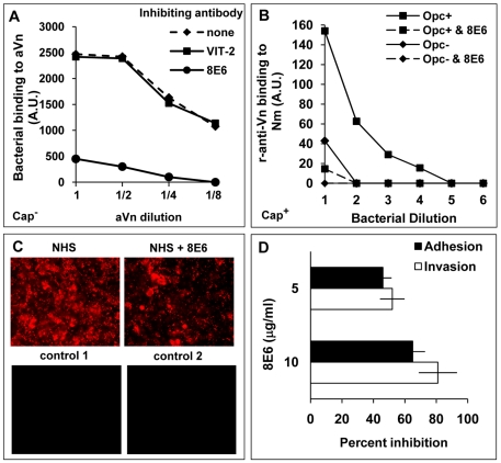
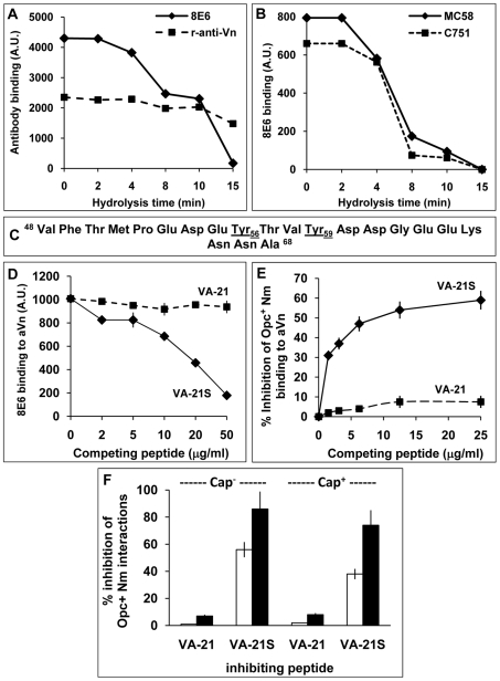
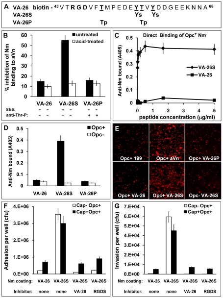
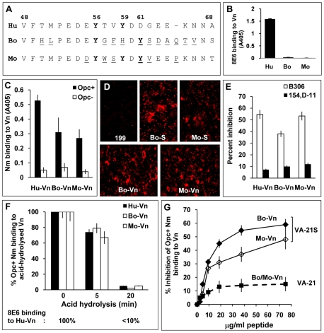
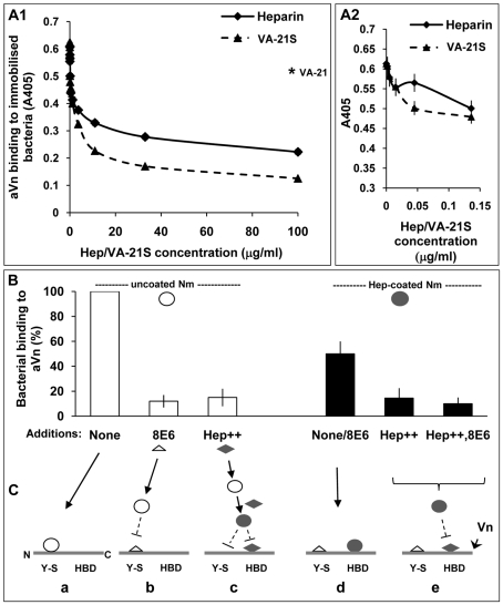
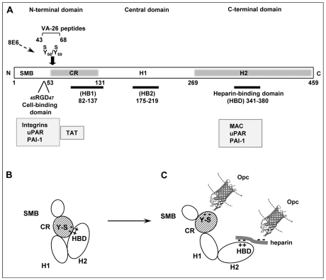
The host vasculature is believed to constitute the principal route of dissemination of Neisseria meningitidis (Nm) throughout the body, resulting in septicaemia and meningitis in susceptible humans. In vitro, the Nm outer membrane protein Opc can enhance cellular entry and exit, utilising serum factors to anchor to endothelial integrins; but the mechanisms of binding to serum factors are poorly characterised. This study demonstrates that Nm Opc expressed in acapsulate as well as capsulate bacteria can increase human brain endothelial cell line (HBMEC) adhesion and entry by first binding to serum vitronectin and, to a lesser extent, fibronectin. This study also demonstrates that Opc binds preferentially to the activated form of human vitronectin, but not to native vitronectin unless the latter is treated to relax its closed conformation. The direct binding of vitronectin occurs at its Connecting Region (CR) requiring sulphated tyrosines Y(56) and Y(59). Accordingly, Opc/vitronectin interaction could be inhibited with a conformation-dependent monoclonal antibody 8E6 that targets the sulphotyrosines, and with synthetic sulphated (but not phosphorylated or unmodified) peptides spanning the vitronectin residues 43-68. Most importantly, the 26-mer sulphated peptide bearing the cell-binding domain (45)RGD(47) was sufficient for efficient meningococcal invasion of HBMECs. To our knowledge, this is the first study describing the binding of a bacterial adhesin to sulphated tyrosines of the host receptor. Our data also show that a single region of Opc is likely to interact with the sulphated regions of both vitronectin and of heparin. As such, in the absence of heparin, Opc-expressing Nm interact directly at the CR but when precoated with heparin, they bind via heparin to the heparin-binding domain of the activated vitronectin, although with a lower affinity than at the CR. Such redundancy suggests the importance of Opc/vitronectin interaction in meningococcal pathogenesis and may enable the bacterium to harness the benefits of the physiological processes in which the host effector molecule participates.


Conflict of interest statement

The authors have declared that no competing interests exist.

Figures

Figure 1
Figure 1. Invasion of HBMECs by capsulate and acapsulate meningococci is dependent on Opc-expression and serum components.
(A) To determine the relative binding of the serum proteins to Opc+ and Opc N. meningitidis, bacteria were incubated with 10% normal human serum (NHS). The binding of vitronectin (Vn) and fibronectin (Fn) was assessed by applying lysates of serum coated, washed bacteria to SDS-polyacrylamide gels and western blotting. Anti-Vn and anti-Fn antibodies followed by AP-conjugated secondary antibodies were used to detect the proteins. Opc-expressing Nm bound to Vn and Fn, whereas Opc-deficient bacteria bound considerably weakly to the proteins. Lane marked NHS was loaded with the serum sample. The two vitronectin bands shown in the left blot correspond to the two molecular forms of vitronectin (75 and 65 kDa) found in human blood, the lower molecular form results from endogenous cleavage of a small region at the C-terminal end of some Vn molecules (see reference 26). (B, C) Relative efficiencies of Opc+ and Opc Nm isolates of strain C751 and MC58 (without or with capsule, pili and Opa expression respectively) in mediating HBMEC adhesion and invasion in serum supplemented media were determined by viable count and gentamicin protection assays. To confirm that gentamicin-resistant bacteria were truly internalised, cytochalasin D (CD) was used in some tests to prevent bacterial uptake into cells. In this case, no bacteria survived the gentamicin treatment (invasion levels indicated by blank columns in C) . (D, E) In similar experiments, various purified serum components were compared with NHS in their ability to support HBMEC adhesion and invasion by acapsulate and capsulate meningococci. When supplemented, the infection media contained 10% NHS or 10 µg/ml of one of the following serum proteins: activated Vn (aVn), native Vn (nVn), cellular Fn (cFn) and plasma Fn (pFn), as in preliminary experiments, these were found to be the optimum concentrations required. The most effective serum component was found to be aVn in enhancing both cellular adhesion as well as invasion. Plasma Fn also supported adhesion and invasion but to a significantly lesser extent (D and E).
Figure 2
Figure 2. Activated vitronectin is adsorbed preferentially from serum by capsulate and acapsulate Opc-expressing N. meningitidis.
(A) To assess which serum protein is preferentially removed from serum by Opc+ Nm, a sample of normal human serum was adsorbed three times sequentially with Opc-expressing phenotype of acapsulate C751. At each stage, aliquots of the adsorbed serum samples were retained. Unadsorbed serum and adsorbed samples were analysed by immuno-dot blot assay (followed by densitometric analysis) for activated and total Vn/Fn levels using the mAb 8E6 and the polyclonal rabbit anti-Vn or anti-fibronectin antibodies. The levels of each of these components extracted from serum by Opc+ Nm as a percent of the total present at the start of extraction are shown. In control experiments, in which Opc Nm were used, 8E6 binding to 3 x adsorbed serum did not alter significantly and amounted to less than 10% loss of aVn (blank column). (B) In a similar experiment, a sample of NHS was adsorbed sequentially with capsulate MC58 Opc+ and Opc phenotypes. The Opc+ bacteria effectively removed a large portion of aVn as assessed by mAb 8E6 binding to serum samples before and after incubation with Nm (B, left). Specific removal of Vn from NHS by Opc+ but not Opc MC58 isolates is apparent from the analysis of serum-adsorbed bacterial pellets which show that bound Vn was only significantly detectable on Opc+ bacteria that were exposed to NHS (B, right).
Figure 3
Figure 3. Deliberate modification of nVn conformation increases Opc–expressing meningococcal interactions with Vn and with HBMECs.
(A) Purified native Vn (nVn) was subjected to limited heat denaturation to relax its folded conformation and reveal the mAb 8E6 binding site. To assess the levels of binding of Opc+ Nm to nVn, heated nVn (h-nVn) and the serum-derived heparin-purified activated Vn preparation (mAb 8E6 binding form), these proteins were overlaid on to immobilised Opc+ and Opc isolates of strain C751 and Vn binding determined using polyclonal anti-Vn antibody. As bacteria lacking Opc exhibited the same level of binding to all vitronectin preparations, these are labelled as (Opc/Vn broken lines). (B) The vitronectin samples used for ELISA were also analysed for their activation status using the mAb 8E6 and the polyclonal anti-Vn antibody (r-anti-Vn) by immuno-dot blotting. The conformation-dependent mAb 8E6 only reacted with aVn and heated nVn demonstrating the exposure of its epitope on heat treatment of nVn. Rabbit antibodies also bound more effectively to the same two forms of Vn suggesting that aVn and h-nVn present larger numbers of epitopes. (C) Immunofluorescence analysis was performed using confluent HBMECs infected with Opc+ and Opc C751 isolates in media supplemented with aVn, nVn and heated nVn illustrating their relative abilities in supporting bacterial adhesion. Bacteria were labelled using anti-Nm antiserum and rhodamine-conjugated secondary antibody.
Figure 4
Figure 4. Monoclonal antibody 8E6 against a cryptic epitope of vitronectin inhibits Opc-mediated interactions of N. meningitidis.
(A) The ability of anti-Vn antibodies 8E6 and VIT-2 to inhibit bacterial binding to immobilised aVn was investigated by immuno-dot blot assays in the presence or absence of the anti-Vn antibodies added at 10 µg/ml prior to Opc+ acapsulate Nm additions. Relative levels of Opc+ Nm binding is shown as arbitrary units determined by densitometric analysis as described in the text. 8E6 but not VIT-2 inhibited direct bacterial binding to purified aVn. (B) 8E6 also prevented adsorption of aVn on to capsulate Opc+ bacteria from NHS; the procedure was carried out as described in the legend to figure 2. The mAb 8E6 when present was used at 10 µg/ml. (C, D) The effect of 8E6 on adhesion/invasion of HBMEC by acapsulate Opc+ bacteria in the presence of 10% NHS was assessed using immunofluorescence adhesion assay (C) or viable count assays (D) performed as described in methods. Bacteria in (C) were labelled using anti-Nm antiserum and rhodamine-conjugated secondary antibody. For controls, either an unrelated primary antibody (control 1) or secondary antibody only (control 2) were used. As addition of 8E6 results in a significant and a dose-dependent inhibition of adhesion and invasion (D), activated Vn appears to be one of the main serum factors supporting Nm interactions with human endothelial cells.
Figure 5
Figure 5. The nature of the vitronectin epitope recognised by N. meningitidis Opc.
(A, B) Acid hydrolysis of vitronectin. Immuno-dot blot assays were performed using acid-treated (1 M HCl, 80°C), immobilised aVn on to nitrocellulose strips. The strips were blocked and overlaid with antibodies or bacteria and analysed for the ability of Vn to retain binding of the mAb 8E6 and polyclonal rabbit anti-Vn antiserum (A) or acapsulate (C751) and capsulate (MC58) Opc+ Nm (B). Binding of bacteria and 8E6 declined simultaneously during acid hydrolysis of Vn known to hydrolyse the sulphate groups. (C–E) Studies using synthetic vitronectin peptides. (C) Sequence of synthetic peptides corresponding to the residues 48–68 containing the 8E6 binding site of vitronectin. The peptide VA-21 was synthesised in a non-sulphated form whereas the peptide named VA-21S was sulphated at residues 56 and 59 as in vitronectin. (D) In a competition immuno-dot blot assay, the ability of the mAb 8E6 to bind to immobilised aVn in the absence or presence of the peptides was assessed. 8E6 binding was quantified using AP-conjugated rabbit anti-mouse secondary antibody followed by densitometry. VA-21S but not VA-21 inhibited 8E6 binding to immobilised aVn. (E) In an ELISA, the binding of acapsulate C751 Opc+ Nm overlaid on to immobilised aVn was inhibited significantly by the addition of increasing concentration of synthetic peptide VA-21S but to a very low extent with the equivalent unsulphated peptide VA-21. (F) To assess the efficacy of VA-21 and VA-21S to inhibit aVn-mediated adhesion and invasion by acapsulate and capsulate Opc+ Nm, viable count experiments were performed. VA-21 or VA-21S peptides (25 µg/ml) were used to pre-coat meningococci (15 min. incubation) which were then added to HBMEC monolayers in infection medium supplemented with aVn (10 µg/ml). VA-21 had no significant effect on adhesion (blank columns) or invasion (filled columns). In contrast, VA-21S reduced cellular adhesion and invasion significantly with a more dramatic effect on invasion levels of both strains.
Figure 6
Figure 6. The VA-26S sulphated peptide contains the features required and sufficient for Opc binding and cellular invasion.
(A) Structures of the biotinylated peptides spanning the Vn region 43–68. VA-26 peptide was unmodified other than at the N-terminal residue which was linked to biotin. Additionally, VA-26S contained sulphated Y56 and Y59 and VA-26P contained phosphorylated T50 and T57. (B) Using 25 µg/ml each of untreated and prior acid-hydrolysed peptides, their relative abilities to inhibit C751 Opc+ bacterial interactions with immobilised aVn were examined by ELISA. Only untreated VA-26S inhibited bacterial binding to aVn. The table summarises the effect of acid-treatment on the peptides (see Figure S1), only the sulphated residues are hydrolysed and consequently only the mAb 8E6 binding to VA-26S is affected after treatment. (C) To examine the direct binding of Opc+ Nm of strain C751 to VA-26S and VA-26, the peptides were first immobilised on extravidin-containing plates (Figure S2). Bacterial binding was detected using anti-Nm antiserum. Opc+ Nm bound significantly only to VA-26S. (D) Relative levels of direct binding of Opc+ and Opc Nm to immobilised VA-26, VA-26S and VA-26P peptides by ELISA. In each case, saturating levels of peptides were used (5 µg/ml each, Figure S2). Bacterial binding as detected using anti-Nm antiserum shows only VA-26S and Opc+ Nm interactions. (E) Immunofluorescence analysis of the ability of VA-26 peptides, which contain the integrin-binding domain RGD, to mediate bacterial binding to HBMECs. Opc+ bacteria but not Opc bacteria localised on to HBMECs in significantly higher numbers in the presence of VA-26S (and aVn control) but not with other peptides (all used at 20 µg/ml). (F and G) To assess the efficacy of VA-26S in mediating acapsulate and capsulate bacterial adhesion and invasion, viable count experiments were performed using VA-26 or VA-26S peptide-coated bacteria. In some experiments VA-26 (20 µg/ml) and RGDS (0.25 mM) were used to preincubate HBMECs to act as inhibitors of RGD-mediated binding of VA-26S-coated bacteria. The data illustrate that two key features (sulphated tyrosines and RGD) present in VA-26S are required and sufficient for mediating bacterial invasion of HBMECs.
Figure 7
Figure 7. Species specificity of Opc and 8E6 and their binding requirements.
(A) Sequences flanking the fully conserved Y56 in human (Hu), bovine (Bo) and murine (mouse, Mo) vitronectins aligned using MegAlign Lasergene software (DNASTAR). The variant residues in bovine and murine vitronectins are underlined. (B) Reactivity of the mAb 8E6 with immobilised vitronectins from distinct sources was analysed by ELISA demonstrating its specificity for human Vn. (C) In a similar experiment, the binding of C751 Opc+ and Opc Nm to immobilised vitronectins was assessed by bacterial overlay. Although human Vn was the most recognised, Opc+ Nm also bound to bovine and mouse Vn at significantly higher levels compared with Opc Nm. (D) The relative abilities of bovine and mouse sera (used at 10%) and purified vitronectins (10 µg/ml) to support C751 Opc+ Nm binding to HBMECs were assessed by immunofluorescence analysis as described in methods. (E) Two mAbs against Opc known to bind to loop 2 (B306) or loops 4/5 (154,D-11) were used in competition studies to assess their ability to block Opc/Vn interactions. Bacterial suspensions were pre-incubated with 30 µg/ml of the antibodies for 20 min prior to addition to Vn. B306 against loop 2 of Opc caused significantly higher inhibition of meningococcal binding to all three Vn samples. (F) The potential role/s of sulphated tyrosines in each of the vitronectins was assessed by acid hydrolysis as described in methods. The relative levels of bacterial binding to the animal vitronectins are shown as blank columns (bovine Vn: middle columns, mouse Vn: right columns) and to human Vn (used for comparison) as filled columns. Percent binding compared with untreated vitronectins demonstrates similar decline in bacterial binding to all three proteins concurrently with the decline in 8E6 binding (monitored simultaneously using human Vn shown below the graph). (G) In a competitive ELISA, VA-21S peptide but not VA-21 inhibited bacterial binding to immobilised bovine and murine vitronectins in a manner similar to human vitronectin suggesting similar mechanisms of targeting the three vitronectins.
Figure 8
Figure 8. Biphasic effect of heparin on bacterial interactions with human vitronectin.
ELISA plates with immobilised acapsulate Opc+ Nm were blocked with BSA block (pre-filtered using heparin-sepharose and DEAE-sephacel) and then overlaid with heparin or VA-21S peptide at the concentrations shown for 20 min prior to the addition of aVn (2.5 µg/ml). Vitronectin binding was assessed as described in Methods. In A1, the results observed over the full range of concentrations are shown whereas in A2 data at a narrower range of heparin concentrations are depicted and VA-21S data are included for comparison. The level of binding in the presence of the control peptide VA-21 when used at a concentration of 100 µg/ml is shown by an asterisk in A1. Data shown are representative of two independent experiments performed using bacteria grown either on GC-agarose or HBHI. Identical results were obtained in both cases. (B) ELISA plates with immobilised aVn were overlaid with uncoated or heparin-precoated bacteria in the absence or presence of excess 8E6 or heparin (Hep++) or both (Hep++,8E6) during the incubation. Bacterial binding was assessed using polyclonal anti-Nm antibody. In each case, percent binding to Vn relative to the highest value observed (i.e. uncoated bacteria in the absence of inhibitors) are shown. (C) The sequence of events consistent with the data in B is represented in a schematic diagram. The horizontal lines represent unfolded Vn molecule (Vn) with its N terminal ‘Y-S’ domain and C-terminally located heparin-binding domain (HBD). These regions may constitute two separate binding sites for Nm on Vn (triangles: mAb 8E6, diamonds: heparin, open circles: uncoated Opc+ Nm, filled circles: heparin-coated Opc+ Nm). a: uncoated Nm bind directly to the Y-S region; b: the mAb 8E6 also binds to Y-S and blocks binding of Nm to the site; c: excess heparin present in the medium coats bacteria and prevents binding to Y-S and in addition, it binds to the HBD and prevents hep-coated Nm binding to HBD; d: Nm precoated with heparin cannot bind to Y-S but in the absence of excess heparin, can bind to HBD; in this case added mAb 8E6 does not affect the level of Nm adhesion to Vn; e: excess free heparin added to the medium binds to HBD and blocks heparin-coated Nm binding to the site; again the presence of mAb 8E6 binding at the CR of Vn has no effect.
Figure 9
Figure 9. Vitronectin domain structure, conformational states and some physiological and bacterial ligand binding sites.
(A) A schematic presentation of linear vitronectin structure showing the positions of the cell-binding motif RGD, sulphated tyrosines (Y56S/Y59S) and the high affinity heparin-binding domain (HBD), the three regions of particular importance in meningococcal interactions with host receptors. The N-terminal domain contains a region homologous to somatomedin B, (SMB, residues 1–44). The central and the C-terminal domains contain regions homologous to haemopexin (H1 and H2). An extended connecting region (CR) joins the N-terminal and the central domains and contains a stretch of acidic residues (53–61) within which two sulphated tyrosines (Y-S) are located. This site is cryptic in the native vitronectin and comprises the mAb 8E6 binding epitope requiring the presence of sulphated residues . The C-terminal domain residues 341–380 contain the main heparin-binding domain (HBD) and are composed of several highly charged residues . Arginine at positions 351/353 may bind directly to heparin . Two other sites implicated in heparin binding (but with low affinity) are shown as HB1 and HB2. Several pathogens bind to the central domain and via heparin to HBD , , . Only Nm has been shown in our current study to bind to the Y-S region in the vitronectin connecting region. Binding positions for physiological ligands are shown (grey boxes). Information is partly based on , , and references there in. (B) An illustration of the folded conformation of the vitronectin molecule which may bring the acidic sulphated tyrosines and the basic HBD in close proximity . (C) Under physiological or in vitro denaturing conditions, Vn structure is relaxed exposing fully the HBD binding site and making available the mAb 8E6 epitope in the CR, both with the capacity to bind to N. meningitidis Opc protein. The binding at the HBD may additionally be assisted by the multimerisation of Vn, which increases the affinity of multimeric Vn for heparin . The molecular model of Opc was kindly provided by Prof. Jeremy Derrick. The Opc crystal structure has been described by Prince et al. ; this study also presents a model of the possible mechanism of interaction of the Opc surface exposed loops with heparin-like proteoglycans.

Similar articles

Cited by

References

    1. Virji M, Makepeace K, Moxon ER. Distinct mechanisms of interactions of Opc-expressing meningococci at apical and basolateral surfaces of human endothelial cells; the role of integrins in apical interactions. Mol Microbiol. 1994;14:173–184. - PubMed
    1. Virji M, Makepeace K, Peak IR, Ferguson DJ, Jennings MP, et al. Opc- and pilus-dependent interactions of meningococci with human endothelial cells: molecular mechanisms and modulation by surface polysaccharides. Mol Microbiol. 1995;18:741–754. - PubMed
    1. Virji M, Makepeace K, Ferguson DJ, Achtman M, Sarkari J, et al. Expression of the Opc protein correlates with invasion of epithelial and endothelial cells by Neisseria meningitidis. Mol Microbiol. 1992;6:2785–2795. - PubMed
    1. Unkmeir A, Latsch K, Dietrich G, Wintermeyer E, Schinke B, et al. Fibronectin mediates Opc-dependent internalization of Neisseria meningitidis in human brain microvascular endothelial cells. Mol Microbiol. 2002;46:933–946. - PubMed
    1. Lambotin M, Hoffmann I, Laran-Chich MP, Nassif X, Couraud PO, et al. Invasion of endothelial cells by Neisseria meningitidis requires cortactin recruitment by a phosphoinositide-3-kinase/Rac1 signalling pathway triggered by the lipo-oligosaccharide. J Cell Sci. 2005;118:3805–3816. - PubMed

Publication types

MeSH terms