. 2010 May 20;5(5):e10659.
doi: 10.1371/journal.pone.0010659.

Alzheimer's disease amyloid-beta links lens and brain pathology in Down syndrome

Affiliations

Alzheimer's disease amyloid-beta links lens and brain pathology in Down syndrome

Juliet A Moncaster et al. PLoS One. .

Abstract

Down syndrome (DS, trisomy 21) is the most common chromosomal disorder and the leading genetic cause of intellectual disability in humans. In DS, triplication of chromosome 21 invariably includes the APP gene (21q21) encoding the Alzheimer's disease (AD) amyloid precursor protein (APP). Triplication of the APP gene accelerates APP expression leading to cerebral accumulation of APP-derived amyloid-beta peptides (Abeta), early-onset AD neuropathology, and age-dependent cognitive sequelae. The DS phenotype complex also includes distinctive early-onset cerulean cataracts of unknown etiology. Previously, we reported increased Abeta accumulation, co-localizing amyloid pathology, and disease-linked supranuclear cataracts in the ocular lenses of subjects with AD. Here, we investigate the hypothesis that related AD-linked Abeta pathology underlies the distinctive lens phenotype associated with DS. Ophthalmological examinations of DS subjects were correlated with phenotypic, histochemical, and biochemical analyses of lenses obtained from DS, AD, and normal control subjects. Evaluation of DS lenses revealed a characteristic pattern of supranuclear opacification accompanied by accelerated supranuclear Abeta accumulation, co-localizing amyloid pathology, and fiber cell cytoplasmic Abeta aggregates (approximately 5 to 50 nm) identical to the lens pathology identified in AD. Peptide sequencing, immunoblot analysis, and ELISA confirmed the identity and increased accumulation of Abeta in DS lenses. Incubation of synthetic Abeta with human lens protein promoted protein aggregation, amyloid formation, and light scattering that recapitulated the molecular pathology and clinical features observed in DS lenses. These results establish the genetic etiology of the distinctive lens phenotype in DS and identify the molecular origin and pathogenic mechanism by which lens pathology is expressed in this common chromosomal disorder. Moreover, these findings confirm increased Abeta accumulation as a key pathogenic determinant linking lens and brain pathology in both DS and AD.


Conflict of interest statement

Competing Interests: Authors Moncaster, Pineda, Lu, Burton, Ghosh, Ericsson, Soscia, Mocofanescu, Folkerth, Robb, Kuszak, Clark, and Hunter report no conflict of interest. The following authors reported consultant activity during the course of this study: Drs. Tanzi and Goldstein, Neuroptix Corp, Acton, MA; Drs. Moir, Tanzi, and Goldstein, Covance, Dedham, MA. No financial support was provided by any commercial entity for this study.

Figures

Figure 1
Figure 1. Phenotypic expression of representative supranuclear cataracts in Down syndrome.
(A-C) Representative supranuclear cataract in a 46-year-old male with Down syndrome observed by slit lamp biomicroscopy. (A) Broad-beam illumination demonstrates numerous cerulean coronary “blue dot” lens opacities. Asterisk denotes first Purkinje image (corneal specular reflection). (B) Retro-illumination reveals a distinctive peripheral ring of opacification in the subequatorial supranuclear subregion of the lens. Red reflex is imparted by retinal reflection. Corneal Purkinje image is evident centrally (asterisk). (C) Inverted grayscale rendering of the retro-illumination image highlights the circumferential subequatorial supranuclear opacification (dashed circle) that characterizes the distinctive Down syndrome cataract phenotype. Corneal Purkinje image is evident centrally (asterisk). (D) Static light scattering intensity plotted as a function of anatomical position within the lens. Axial location is referenced to the dashed line and anatomical orientation axes represented in the inset. (E-H) Representative stereophotomicroscopic images of ex vivo lenses from subjects with Down syndrome of indicated ages and gender. Colored dots indicate superior and inferior extent of the slit beam ribbon defining the posterior surface of each imaged lens. White dots indicate superior and inferior extent of the light reflex on the anterior aspect of each imaged lens. (E) Lens from a 2-year-old male with Down syndrome. Slit beam photomicroscopy demonstrates a grossly normal transparent lens. (F) Lens from a 42-year-old female with Down syndrome. Supranuclear opacification is evident in a semicircular arc (dashes) with prominent cortical spokes and moderate lenticular brunescence. (G) Lens from a 61-year-old female with Down syndrome. Distinctive supranuclear opacification describes an incomplete supranuclear arc (dashes). Cortical spokes and moderate lenticular brunescence are present. (H) Lens from a 64-year-old male with Down syndrome. Prominent circumferential opacification is present as a subequatorial supranuclear ring cataract (dashed circle). This cataract describes an annular half torroid that follows the anteroposterior orientation of the supranuclear fiber cells enveloping the peripheral extent of the embryonic nucleus. Cortical spokes and moderate lenticular brunescence are evident. This same lens is presented as a stereo image pair with and without intact zonule fibers (Fig. S1 and Fig. 2, respectively). See text for details.
Figure 2
Figure 2. Stereo images demonstrating mature supranuclear lens pathology in Down syndrome and Alzheimer's Disease.
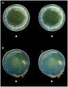
(A) Characteristic circumferential supranuclear cataract in the lens of a 64-year-old male subject with Down syndrome. This distinctive cataract is evident as an annular half-toroid band of opacification in the deep cortical and supranuclear subregions of the lens. This same lens specimen is presented as a slit lamp biomicrocopic image (Fig. 1H) and as a stereo image pair (with intact zonule fibers, Fig. S1). This dramatic Down syndrome cataract is phenotypically comparable to the incomplete subequatorial supranuclear cataract observed in the lens of a 76-year-old male subject with advanced Alzheimer's disease (B). These distinctive supranuclear cataracts are not observed in age-normal control subjects. See text for details.
Figure 3
Figure 3. Alzheimer's disease amyloid-β (Aβ) pathology in Down syndrome lens.
(A) Histological section of a human lens obtained from a 21-year-old male subject with Down syndrome. Hematoxylin and eosin. (B) Schematic diagram identifying anatomical regions of the human lens (adapted from Histology of the Human Eye: An Atlas and Textbook [52]). (C) Archival rendering of classical coronary cerulean “flake” arcuate cataracts in a 40-year-old male with presumptive Down syndrome (from Lowe, 1949 [23]). This historical folio drawing illustrates the mature Down syndrome supranuclear cataract phenotype with dominant subequatorial localization and anteroposterior extension. (D-F) Congo red amyloid histochemical analysis of lenses from a 21-year-old male with Down syndrome and age-matched normal male control. (D) Congophilia in the cortex and supranuclear subregion of the lens from a 21-year-old male with Down syndrome. (E) Intense co-localizing apple-green birefringence in the corresponding cortical and supranuclear subregions of the same Congo red-stained Down syndrome lens imaged with cross-polarized illumination. (F) Amyloid histochemical analysis of a lens from a 21-year-old normal control subject did not demonstrate Congophilia nor classical apple-green birefringence under identical cross-polarized illumination. (G) Aβ immunoreactivity in the epithelium, deep cortex, and supranuclear regions in a lens from a 22-year-old male with Down syndrome. Inset, magnified detail of the anterior lens (box). Cap, capsule; epi, epithelium; cor, cortex; snc, supranucleus; nuc, nucleus. (H) Confirmation of anti-Aβ antibody specificity in the same Down syndrome lens by immunodepletion of the detection antibody with synthetic human Aβ. Adjacent section of the same Down syndrome lens in Fig. 3G (I) Absence of Aβ immunoreactivity in the lens of a normal 22-year-old male control subject. (J) Anti-Aβ immunogold electron microscopic analysis of lens from a 58-year-old male with Down syndrome. Heterogeneously distributed anti-Aβ immunoreactive protein aggregates of dimensions ∼5–200 nm localize heterogeneously within the lens fiber cell cytoplasm. Aβ immunoreactivity was not detected at the plasmalemma. Boxed region denotes magnified area shown in Fig. 3k . Scale bar = 500 nm. (K) High-magnification electron micrograph of a single Aβ-immunoreactive cytoplasmic protein aggregate (arrow) in a lens fiber cell from same Down syndrome lens section shown in Fig. 3J. Multiple immunogold particles detect a single cytoplasmic protein aggregate with longest axial cross-section ∼50 nm. Scale bar = 50 nm. (L) Confirmation of anti-Aβ antibody specificity. Anti-Aβ immunostaining was not detected in Down syndrome lens when probed with immunodepleted anti-Aβ antibody.
Figure 4
Figure 4. Peptide sequencing of human Aβ from Down syndrome lens.
Tryptic digest tandem mass spectrometry sequencing of a ∼4 kDa HPLC eluate derived from human Down syndrome lens protein extract. The retention time of the immunopurified HPLC eluate used for sequencing was identical to synthetic human Aβ. The red shade box denotes the detected 12-residue internal tryptic peptide sequence 17LVFFAEDVGSNK28 that uniquely identifies human Aβ. We detected a second unique tryptic peptide 6HDSGYEVHHQK16 in another analysis (purple underline).
Figure 5
Figure 5. Lens Aβ extraction efficiency in sodium dodecyl sulfate and formic acid.
See text for details.
Figure 6
Figure 6. Increased Aβ expression in Down syndrome lens and brain.
(A-D) Anti-Aβ ELISA analysis of human lens (A) and brain (B) fractionated by Aβ isoform. (A) Lens homogenates demonstrate elevated lens Aβ in subjects with Down syndrome (n = 8; p = 0.027) compared to normal controls (n = 15). (B) Brain homogenates demonstrate elevated Aβ in subjects with Down syndrome (n = 8; p = 0.007) and Alzheimer's disease (n = 6; p = 0.026) compared to normal controls (n = 6). (C) Immunoblot analysis of Down syndrome lens and brain homogenates reveals an intense Aβ-immunoreactive band that migrated with an apparent molecular weight of ∼4kDa corresponding to synthetic human Aβ monomer (arrow). Note strong band corresponding to Aβ monomer in the 2-year-old Down syndrome lens (lane 7) and apparent shift from lower- to higher-order oligomeric Aβ in Down syndrome lenses with advancing age (lanes 7–9). Protein loading was normalized in each lane.
Figure 7
Figure 7. Aβ potentiates lens protein aggregation and Rayleigh light scattering in vitro.
(A) Brightfield photomicroscopic image of a Congo red-stained protein aggregate (arrow) formed during incubation of human lens protein extract with synthetic human Aβ. (B) Same Congo red-stained lens protein aggregate demonstrating classical amyloid birefringence under cross-polarized illumination. (C) Anti-Aβ/anti-αB-crystallin double immunogold electron microscopic analysis of protein aggregates formed during incubation of human lens protein with synthetic human Aβ. Larger immunogold particles (15 nm diameter) detect Aβ. Smaller immunogold particles (10 nm diameter) detect αB-crystallin. Box indicates region of detail highlighted in Fig. 7D . Scale bar = 200 nm. Inset, hetero-oligomeric composition noted in a single protein aggregate demonstrated by double immunogold (anti-Aβ and anti-αB-crystallin) electron microscopy. Analysis was performed on human lens protein incubated with synthetic human Aβ. Larger immunogold particles (15 nm diameter) detect Aβ (black arrow). Smaller immunogold particles (10 nm diameter) detect αB-crystallin (white arrow). Scale bar = 50 nm. (D) Quasi-elastic light scattering (QLS) analysis detects increased backscattered light due to concentration-and time-dependent protein aggregation during incubation of human lens protein with synthetic human Aβ. Experimental samples studied by QLS were analyzed by amyloid histochemistry (Fig. 7A,B) and double (anti-Aβ and anti-αB-crystallin) immunogold electron microscopy (Fig. 7C).
Figure 8
Figure 8. Age-dependent supranuclear cataractogenesis in Down syndrome.
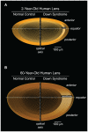
The anatomical localization of the characteristic Down syndrome cataract phenotype (white shading) reflects the temporal origin and natural history of the underlying lens pathology. Fetal lens fiber cells are not involved in this pathogenic process. Parentheses indicate equatorial axial extent of age-dependent disease-linked Aβpathology in the supranuclear subregion of Down syndrome lenses. See text for details.
Figure 9
Figure 9. Model pathogenic pathways in Down syndrome brain and lens.
Triplication of human chromosome 21 in Down syndrome results in increased dosage of the APP gene (21q21), overexpression of the Alzheimer's disease amyloid-β precursor protein (APP), and progressive accumulation of amyloidogenic amyloid-β peptides (Aβ) in the brain and lens. Deposition of Aβ in both anatomical compartments results in age-dependent Aβ amyloid pathology and disease-linked tissue-specific phenotypes in both Down syndrome and Alzheimer's disease. See text for details.

Similar articles

Cited by

References

    1. Down JLH. Observations on an ethnic classification of idiots. London Hosp Clin Lect Rep. 1866;3:259.
    1. Epstein CJ. Down syndrome, trisomy 21. In: Scriver CR, Beaudet AL, Sly WS, Valle D, editors. Metabolic Basis of Inherited Disease. New York: McGraw-Hill; 1989. pp. 291–326.
    1. Online Mendelian Inheritance in Man - Down Syndrome. Available: http://www.ncbi.nlm.nih.gov/entrez/dispomim.cgi?id=190685. Accessed 25 April 2010.
    1. Pueschel SM. Clinical aspects of Down syndrome from infancy to adulthood. Am J Med Genet. 1990;Suppl 7:52–56. - PubMed
    1. Roizen NJ, Patterson D. Down's syndrome. The Lancet. 2003;361:1281–1289. - PubMed

Publication types

MeSH terms