. 2010 May 20;6(5):e1000956.
doi: 10.1371/journal.pgen.1000956.

Mutation in archain 1, a subunit of COPI coatomer complex, causes diluted coat color and Purkinje cell degeneration

Affiliations

Mutation in archain 1, a subunit of COPI coatomer complex, causes diluted coat color and Purkinje cell degeneration

Xinjie Xu et al. PLoS Genet. .

Abstract

Intracellular trafficking is critical for delivering molecules and organelles to their proper destinations to carry out normal cellular functions. Disruption of intracellular trafficking has been implicated in the pathogenesis of various neurodegenerative disorders. In addition, a number of genes involved in vesicle/organelle trafficking are also essential for pigmentation, and loss of those genes is often associated with mouse coat-color dilution and human hypopigmentary disorders. Hence, we postulated that screening for mouse mutants with both neurological defects and coat-color dilution will help identify additional factors associated with intracellular trafficking in neuronal cells. In this study, we characterized a mouse mutant with a unique N-ethyl-N-nitrosourea (ENU)-induced mutation, named nur17. nur17 mutant mice exhibit both coat-color dilution and ataxia due to Purkinje cell degeneration in the cerebellum. By positional cloning, we identified that the nur17 mouse carries a T-to-C missense mutation in archain 1 (Arcn1) gene which encodes the delta subunit of the coat protein I (COPI) complex required for intracellular trafficking. Consistent with this function, we found that intracellular trafficking is disrupted in nur17 melanocytes. Moreover, the nur17 mutation leads to common characteristics of neurodegenerative disorders such as abnormal protein accumulation, ER stress, and neurofibrillary tangles. Our study documents for the first time the physiological consequences of the impairment of the ARCN1 function in the whole animal and demonstrates a direct association between ARCN1 and neurodegeneration.


Conflict of interest statement

The authors have declared that no competing interests exist.

Figures

Figure 1
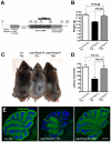
Figure 1. Coat color dilution and Purkinje cell degeneration in nur17 mice.
(A) Diluted coat color in nur17 mice (right) compared with control mice (left). (B) Body weight of 2-month old nur17 (n = 7, female) and control (n = 5, female) mice. Error bars represent standard error. **p<0.01 (C) Pigment distribution in the hair of control and nur17 mice. Base, midshaft and tips of the hair of control (top) and nur17 (bottom) mice are shown. Representative pictures from 30 hairs of each genotype are shown. (D) Ratio of hairs with abnormal pigmentation in the base (left), midshaft (center) and tip (right). Total of 150 hairs from 5 nur17 and 5 control mice (30 hairs each) were examined. Error bar represent standard error. *p<0.05; **p<0.01 (E) Degenerative loss of the Purkinje cells in nur17 mice. Purkinje cells are labeled with anti-calbindin (green) and nuclei are stained with DAPI (blue). Note that by 2 months of age, nur17 mice lose their Purkinje cells in Lobule IX. Upper scale bar: 1mm; Bottom scale bar: 0.1mm.
Figure 2
Figure 2. Positional cloning of the nur17 gene.
(A) The haplotypes for 822 chromosomes from 411 F2 mice are indicated. (B) Genotypes and phenotypes of two recombinant mice that were critical for determining the minimal region for the nur17 locus. (C) Minimal genetic region of the nur17 locus. (D) Point mutation in the Arcn1 gene from nur17 mice. T to C nucleotide transition causes an amino-acid change from Ile to Thr at amino acid (aa) 422. (E) Point mutation is located in the 10th exon of the Arcn1 gene in nur17 mice.
Figure 3
Figure 3. Phenotypes of nur17 mice are rescued by wild-type Arcn1.
(A) Transgenic construct for generating the transgenic mice expressing Arcn1 gene. Restriction enzyme (PstI) sites to digest DNA for southern blotting were labeled. The black bar represents the region used for the probe for southern blot. (B) Rescue of smaller body weight by transgene in nur17 mice. Smaller size in nur17 mice (middle, n = 13, female) is rescued by the introduction of transgene, Arcn1 (right, n = 6, female) (p = 0.003). There is no significant difference between the weight of rescued mice (right) and the control mice (left, n = 7, female) (p = 0.44). (C) Rescue of coat color dilution. Coat color dilution phenotype in nur17 mice (middle) is rescued by the introduction of transgene, Arcn1 (right). (D) Rescue of the motor coordination defect. Latency to fall on the accelerating rotatod was recorded for control mice (n = 5), nur17 mice (n = 5) and nur17 mice with the Arcn1 transgene (n = 5). The rotarod performance was significantly improved by the transgene (p = 0.015), and was comparable to that in control mice (p = 0.613). (E) Rescue of Purkinje cell degeneration. Purkinje Cell degeneration phenotype in 2-month old nur17 mice (middle) is completely rescued by the introduction of transgene, Arcn1 (right). Purkinje cells are labeled with anti-calbindin (green) and nuclei are stained with DAPI (blue). Scale bar: 1mm.
Figure 4
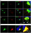
Figure 4. Subcellular localization of ARCN1.
(A) Primary culture of wild-type mouse melanocytes are labeled with anti-ARCN1 (green) indicating the localization of ARCN1 and anti-Golgi protein (red) (i); anti-KDEL (red) (ii) and anti-βCOP (red) (iii). (B) Neuro-2a cells transfected with constructs for GFP-tagged ARCN1 (green) and V5-tagged βCOP (red). Nuclei are stained with DAPI (blue). Merged pictures are shown and high magnification pictures are shown in the right panel. Scale bar for low magnification: 10µm. Scale bar for high magnification: 5µm.
Figure 5
Figure 5. Kinetic analysis of biosynthesis and transport of Tyrp1 in the melanocyte.
(A) Pulse chase analysis of control and nur17 melanocytes. Control and nur17 melanocytes were pulse labeled for 10 min with [35S] methionine followed by a chase as the indicated times. The size of Tryp1 is monitored after pulse labeling with [35S] and analyzed by 9% SDS-PAGE. (Molecular weight markers are shown on the left.) (B) Quantification of the proportion of Tyrp1 without complex N-linked glycans at 5 different chasing time points (0min, 15min, 25min, 35min and 45min). Error bar represents standard error.
Figure 6
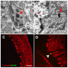
Figure 6. Abnormal protein aggregation and upregulated ER stress marker in the cerebellum of nur17 mice.
(A) Ultrastructural analysis showed protein inclusion (red arrow) in the dendrite of PC. (B) Protein inclusion (red arrow) also exists in the perinuclear region of PC. PC nucleus is marked by a red star. (C, D) PC labeled with anti-Calbindin (red) is negative for anti-CHOP antibody staining (green) in control mice (C) but positive in nur17 mice (D, white arrowhead). Scale bar: 30um.
Figure 7
Figure 7. Neurofibrilary tangles in the cerebellum of nur17 mice.
(A, B) Ultrastructural analysis showed abnormal filamentous lesions (red arrowhead) in the cerebellum of nur17 mice (A), while such lesions are not observed in control mice (B). The inset in (A) shows the higher magnification of filamentous lesion. (C–E) Gallyas staining for control (C) and nur17 mice (D, E). Arrow denotes the PC that is positive for Gallyas staining. Scale bar for (C, D): 1mm. Scale bar for (E): 0.1mm.

References

    1. Schwartz AL. Cell biology of intracellular protein trafficking. Annu Rev Immunol. 1990;8:195–229. - PubMed
    1. Annaert W, De Strooper B. A cell biological perspective on Alzheimer's disease. Annu Rev Cell Dev Biol. 2002;18:25–51. - PubMed
    1. Uemura K, Kuzuya A, Shimohama S. Protein trafficking and Alzheimer's disease. Curr Alzheimer Res. 2004;1:1–10. - PubMed
    1. DiFiglia M, Sapp E, Chase K, Schwarz C, Meloni A, et al. Huntingtin is a cytoplasmic protein associated with vesicles in human and rat brain neurons. Neuron. 1995;14:1075–1081. - PubMed
    1. Gil JM, Rego AC. Mechanisms of neurodegeneration in Huntington's disease. Eur J Neurosci. 2008;27:2803–2820. - PubMed

Publication types