. 2010 May 20;6(5):e1000964.
doi: 10.1371/journal.pgen.1000964.

FACT prevents the accumulation of free histones evicted from transcribed chromatin and a subsequent cell cycle delay in G1

Affiliations

FACT prevents the accumulation of free histones evicted from transcribed chromatin and a subsequent cell cycle delay in G1

Macarena Morillo-Huesca et al. PLoS Genet. .

Abstract

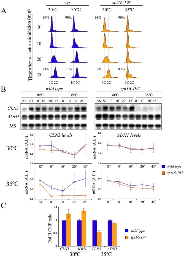
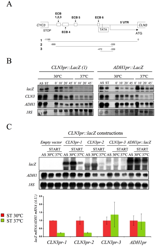
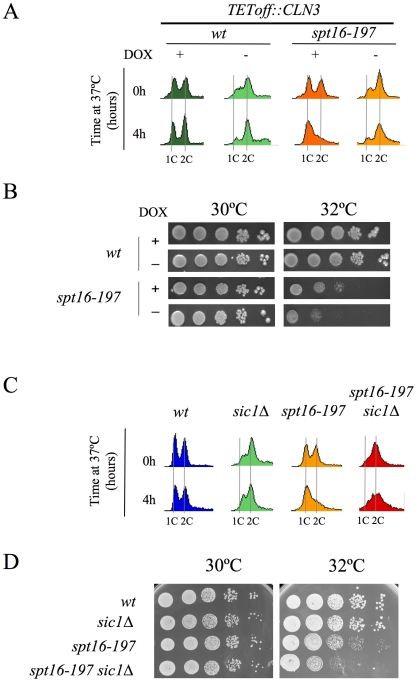
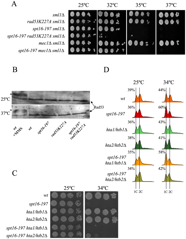
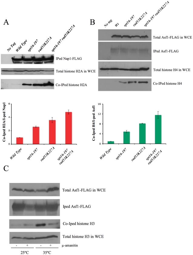
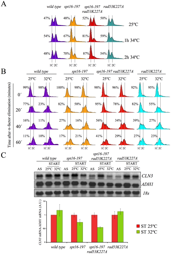
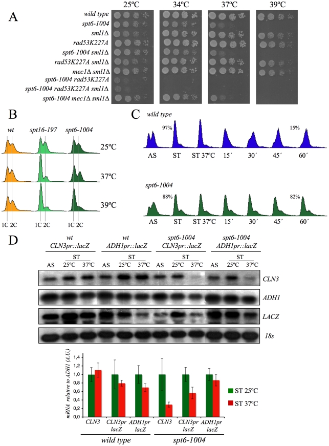
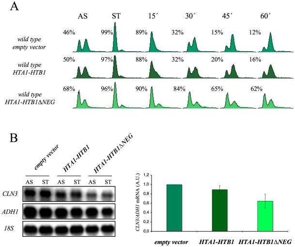
The FACT complex participates in chromatin assembly and disassembly during transcription elongation. The yeast mutants affected in the SPT16 gene, which encodes one of the FACT subunits, alter the expression of G1 cyclins and exhibit defects in the G1/S transition. Here we show that the dysfunction of chromatin reassembly factors, like FACT or Spt6, down-regulates the expression of the gene encoding the cyclin that modulates the G1 length (CLN3) in START by specifically triggering the repression of its promoter. The G1 delay undergone by spt16 mutants is not mediated by the DNA-damage checkpoint, although the mutation of RAD53, which is otherwise involved in histone degradation, enhances the cell-cycle defects of spt16-197. We reveal how FACT dysfunction triggers an accumulation of free histones evicted from transcribed chromatin. This accumulation is enhanced in a rad53 background and leads to a delay in G1. Consistently, we show that the overexpression of histones in wild-type cells down-regulates CLN3 in START and causes a delay in G1. Our work shows that chromatin reassembly factors are essential players in controlling the free histones potentially released from transcribed chromatin and describes a new cell cycle phenomenon that allows cells to respond to excess histones before starting DNA replication.


Conflict of interest statement

The authors have declared that no competing interests exist.

Figures

Figure 1
Figure 1. CLN3 expression is down-regulated after Spt16 inactivation.
Wild-type (FY120) and spt16-197 (FY348) cells grown asynchronously (AS) were synchronized at START by treatment with alpha-factor for two hours at 30°C (ST), followed by one additional one hour at 30°C or 35°C in the presence of the mating pheromone. Cells were then released from the arrest at time 0 at either 30°C or 35°C by washing out the alpha-factor. Samples were taken at different time points to analyze the DNA content by flow cytometry and the proportion of unbudded cells by microscopy (A), and to quantify the mRNA levels of the indicated genes by Northern blot analysis (B). Transcripts levels were represented as arbitrary units (AU) after normalization with 18S rRNA. The results of a typical experiment and the quantification of three independent experiments are shown. (C) Relative RNApol II binding to the transcribed region of CLN3 and ADH1 in spt16-197. Cells were synchronized with alpha-factor for two hours at 30°C, followed by one additional one hour at 30°C or 35°C in the presence of the mating pheromone. The 8WG16 antibody, recognizing the CTD repeats of Rpb1, was used.
Figure 2
Figure 2. The CLN3 promoter mediated the down-regulation of its mRNA levels after Spt16 inactivation.
(A) Diagram of the plasmid-borne lacZ fusions, encoding E. coli ß-galactosidase, that were generated by introducing different fragments of the CLN3 promoter region. Constructions were numbered 1 to 3. The positions of the early cell cycle boxes (ECB) and the TATA box are indicated. An ADH1 promoter-lacZ fusion was used as a constitutively expressed control. (B) The mRNA levels of CLN3pr::lacZ and ADH1pr::lacZ fusion. The spt16-197 (FY348) strain, transformed with the plasmids bearing lacZ fusion number 1 (CLN3pr::lacZ) or ADH1pr::lacZ, was grown until the mid-log phase (AS), and was then synchronized at START (ST) by alpha-factor treatment for 2h at 30°C, followed by one additional hour of incubation at 30°C or 37°C in the presence of the mating pheromone. Cells were then transferred to a fresh medium without alpha-factor (time 0h) to allow cells to progress at 30°C or 37°C. At the indicated times, samples were taken to analyze the transcript levels by Northern blot analysis with the indicated probes. The results of a typical experiment are shown. (C) The spt16-197 (FY348) cells were transformed with the plasmids bearing the three lacZ fusions described in (A) or ADH1pr::lacZ. These transformants were grown until the mid-log phase (AS) and were synchronized with alpha-factor at 30°C (START), followed by an additional hour at 30°C or 37°C in the presence of the mating pheromone, as indicated. The transcript levels were quantified by Northern blot analysis. The results of a typical experiment and the quantification of three independent experiments are shown. The CLN3 levels were normalized to ADH1. AU: arbitrary units.
Figure 3
Figure 3. The forced entry of Spt16-deficient cells into the S-phase lowered their viability.
(A) Wild-type (FY120) and spt16-197 (FY348) cells, containing the centromeric pTetoff-CLN3-HA plasmid, were grown in a synthetic medium (SC without uracil) at 30°C in the presence (+) or absence (−) of doxycycline (5µg/ml). Exponentially growing cells were then transferred to 37°C for four hours and analyzed by flow cytometry. (B) The wild-type and spt16-197 cells containing the pTetoff-CLN3-HA plasmid were grown until the mid-log phase in a synthetic medium (SC-URA) at 30°C in the presence (+) or absence (−) of doxycycline. Then, 10-fold serial dilutions were spotted onto the same media and incubated at the permissive (30°C) or semi-permissive (32°C) temperature. The first spot on the left corresponds to 5 µl of the undiluted culture. Pictures correspond to three days of incubation. No growth was detected on the forth spot of spt16-197 minus doxycycline at 32°C, even after five days of incubation. (C) The wild-type (MMY18.10), sic1Δ (MMY18.12), spt16-197 (MMY18.11) and spt16-19 sic1Δ (MMY18.9) cells growing exponentially in YPD at 30°C were shifted to 37°C for four hours. Cells were then analyzed by flow cytometry. (D) The exponentially growing cells from the strains mentioned in (C) were spotted onto YPD plates, as described in (B), and incubated at 30°C and 32°C as indicated. Pictures correspond to three days of incubation. No growth was detected on the fifth spot of the double mutant at 32°C, even after five days of incubation.
Figure 4
Figure 4. Genetic interactions connect FACT dysfunction to free histones.
(A) rad53K227A enhances the thermosensitivity of spt16-197, irrespectively of the DNA damage checkpoint. Strains VO1-1, VO1-2, VO1-3, VO1-5, VO1-6, and VO1-8 were grown in YPD medium at 25°C. 10-fold serial dilutions were plated on YPD plates and incubated for 3 days at the indicated temperatures. (B) FACT dysfunction did not induce the hyperphosphorylation of Rad53p. The indicated strains (FY120, FY348, VO3, and VO7) were grown in YPD at either 25°C or 37°C for two hours. The TCA-treated protein extracts were analyzed by Western blot analysis with the goat anti-Rad53 polyclonal antibody. The hyperphosphorylation of Rad53 (shown by an *) was evidenced in the wild-type cells treated with 0.02% MMS. (C) hta2/htb2Δ partially suppressed spt16-197. Strains (FY120, FY348, FY710, DMY10, DMY11, and DMY12) were grown in YPD medium at 25°C. 10-fold serial dilutions of the indicated strain were plated on YPD plates and incubated for three days at the indicated temperatures. (D) Cells of strains FY120 (WT), FY348 (spt16-197), FY710 (hta1Δ–htb1Δ), DMY10 (hta2Δ–htb2Δ), DMY11 (spt16-197 hta1Δ–htb1Δ), and DMY12 (spt16-197 hta2Δ–htb2Δ), exponentially growing in YPD at 25°C, were shifted to 37°C for four hours or kept at 25°C. Samples were taken to analyze the DNA content by flow cytometry. Numbers indicate the proportion of G1 cells.
Figure 5
Figure 5. FACT dysfunction causes a transcription-dependent accumulation of free histones.
(A) Histone H2A levels associated with chaperone Nap1. The indicated strains carrying Nap1-FLAG were grown exponentially at 25°C and then shifted for two hours to 37°C, prior to harvesting the cells for whole cell extract (WCE) preparation. Co-immunoprecipitation assays were carried out as described in the Materials and Methods. The levels of immunoprecipitated (IPed) Nap1 and co-immunoprecipitated (co-IPed) H2A are indicated. The histogram shows the relative accumulation of H2A on Nap1 compared to the wild-type cells (the data have been normalized to the amount of Nap1 actually IPed). The total levels of H2A in WCE are shown to demonstrate that roughly equal amounts of WCE were used for the immunoprecipitation reactions. (B) The histone H4 levels associated with Asf1 in the spt16-197 cells. The indicated strains carrying FLAG-tagged Asf1 were grown in YPD media at 25°C, while the exponentially growing cells were harvested for WCE preparation. The co-immunoprecipitation assays for Asf1-H4 were carried out as indicated in the Materials and Methods. The total histone H4 and the total Asf1-FLAG levels in the WCE are shown to demonstrate that roughly equal amounts of WCE were used for the immunoprecipitation reactions. The histogram displays the relative accumulation of H4 on Asf1 if compared to the wild-type cells (the data have been normalized to the amount of Asf1 actually IPed). (C) The accumulation of the free histones in G1 upon FACT dysfunction is dependent on transcription. The spt16-197 cells (FY348) carrying FLAG-tagged Asf1 were grown in YPD at 25°C, alpha factor-treated for two hours at 25°C, divided into four equal aliquots, and finally incubated with or without alpha-amanitin for two hours at 25°C or 35°C in the continued presence of the pheromone. Following this, cells were harvested and processed as described for (A). The Asf1-FLAG and histone H3 levels in WCE were shown to demonstrate that roughly equal amounts of WCE were used for the immunoprecipitation reactions.
Figure 6
Figure 6. Excess histones induce a cell cycle delay in G1.
(A) The spt16-197 (FY348) and spt16-197 rad53K227A (VO7) cells growing exponentially in YPD at 25°C were shifted to 32°C at the indicated times. DNA content was analyzed by flow cytometry and the proportion of unbudded cells was quantified by microscopy. (B) The wild-type (FY120), spt16-197 (FY348), spt16-197 rad53K227A (VO7) and rad53-K227A (V03) cells were synchronized at START by a treatment with alpha-factor for two hours at 25°C, followed by an additional one hour at 25°C or 32°C in the presence of mating pheromone. Cells were then released from the G1-arrest at time 0 at either 25°C or 32°C by washing out the alpha-factor. Samples were taken at the different time points to analyze the DNA content by flow cytometry and to measure the proportion of unbudded cells by microscopy. (C) Those samples from asynchronous cultures and from the time 0 of B were taken to analyze the CLN3 mRNA levels by Northern blot. The results of a significant experiment and the average quantification of three independent experiments are shown. The CLN3 mRNA levels were normalized to ADH1.
Figure 7
Figure 7. Spt6 dysfunction provokes G1/S defects and lowers cell viability in combination with rad53K227A.
(A) Strains FY120, FY2180, VO1, VO3, DMY5, VO4, VO2, DMY6, DMY7, and DMY8 were grown in YPD at 25°C, spotted onto YPD plates and incubated for three days at the indicated temperature. (B) The wild-type (FY120), spt16-197 (FY348), and spt6-1004 (FY2180) cells exponentially growing in YPD at 25°C, were shifted for two hours at the indicated temperatures. Samples were then taken to analyze the DNA content by flow cytometry. (C) The wild-type (FY120) and spt6-1004 (FY2180) cells were synchronized at START by alpha-factor treatment for 2h at 25°C, followed by an additional one hour at 37°C in the presence of the mating pheromone. Cells were then released from the G1-arrest at time 0 at 37°C by washing out the alpha-factor. Samples were taken at the different time points to analyze the DNA content by flow cytometry and to measure the proportion of unbudded cells by microscopy. (D) The mRNA levels of CLN3 and CLN3pr::lacZ in spt6-1004. The wild-type (FY120) and spt6-1004 (FY2180) cells were transformed with the plasmids bearing the CLN3pr::lacZ fusion number 1 (see Figure 2A) or ADH1pr::lacZ. These transformants were grown until the mid-log phase (AS) and were alpha factor-synchronyzed at 25°C (START), followed by one additional hour at 37°C in the continued presence of pheromone. The RNA samples were taken to analyze the transcript levels. mRNAs were quantified by Northern blot analysis. The results of a typical experiment and the average quantification of three independent experiments are shown. The CLN3 mRNA levels were normalized to ADH1, and the values of each strain at 37°C were represented in relation to 25°C.
Figure 8
Figure 8. The overexpression of histones in the wild-type cells induces G1 delay.
(A) Wild-type cells were transformed with pRS316 (empty vector) or with the analogous centromeric plasmids expressing either HTA1–HTB1 or a mutant version of this locus lacking the sequence that mediates its transcriptional repression in response to the free histones (HTA1–HTB1ΔNEG). The transformants were grown exponentially in a selective medium (AS), synchronized at START by treatment with alpha-factor for two hours (ST) and released from the arrest by washing out the alpha-factor. Samples were then taken at the different time points to analyze the DNA content by flow cytometry and the proportion of unbudded cells by microscopy. (B) Aliquots from the synchronized cells used in (A) were taken to analyze the CLN3 mRNA levels in START by Northern blot. The results of a typical experiment and the average quantification of three independent experiments are shown. The CLN3 mRNA levels have been normalized to ADH1.

Similar articles

Cited by

References

    1. Reinberg D, Sims RJ., 3rd de FACTo nucleosome dynamics. J Biol Chem. 2006;281:23297–23301. - PubMed
    1. Formosa T. FACT and the reorganized nucleosome. Mol Biosyst. 2008;4:1085–1093. - PubMed
    1. Orphanides G, LeRoy G, Chang CH, Luse DS, Reinberg D. FACT, a factor that facilitates transcript elongation through nucleosomes. Cell. 1998;92:105–116. - PubMed
    1. Pavri R, Zhu B, Li G, Trojer P, Mandal S, et al. Histone H2B monoubiquitination functions cooperatively with FACT to regulate elongation by RNA polymerase II. Cell. 2006;125:703–717. - PubMed
    1. Jimeno-Gonzalez S, Gomez-Herreros F, Alepuz PM, Chavez S. A gene-specific requirement for FACT during transcription is related to the chromatin organization of the transcribed region. Mol Cell Biol. 2006;26:8710–8721. - PMC - PubMed

Publication types

MeSH terms