. 2010 May 20;6(5):e1000915.
doi: 10.1371/journal.ppat.1000915.

EBV promotes human CD8 NKT cell development

Affiliations

EBV promotes human CD8 NKT cell development

Yuling He et al. PLoS Pathog. .

Retraction in

  • Retraction: EBV promotes human CD8 NKT cell development.
    Yuling H, Ruijing X, Xiang J, Li L, Lang C, Jie X, Wei X, Yujuan W, Lijun Z, Rui Z, Xinti T, Yongyi B, Yan-Ping J, Youxin J, Jinquan T. Yuling H, et al. PLoS Pathog. 2010 Dec 3;6(12):10.1371/annotation/1730f52d-2ba9-4f08-b330-47d71b31ae4a. doi: 10.1371/annotation/1730f52d-2ba9-4f08-b330-47d71b31ae4a. PLoS Pathog. 2010. PMID: 21151944 Free PMC article. No abstract available.

Abstract

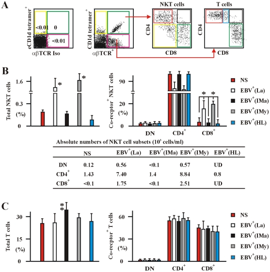
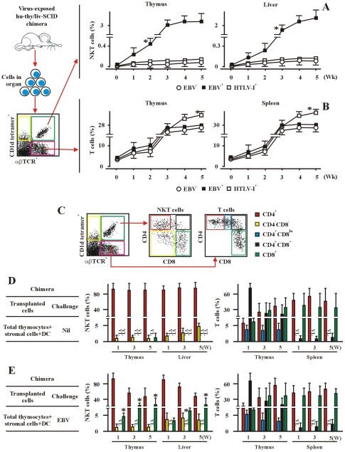
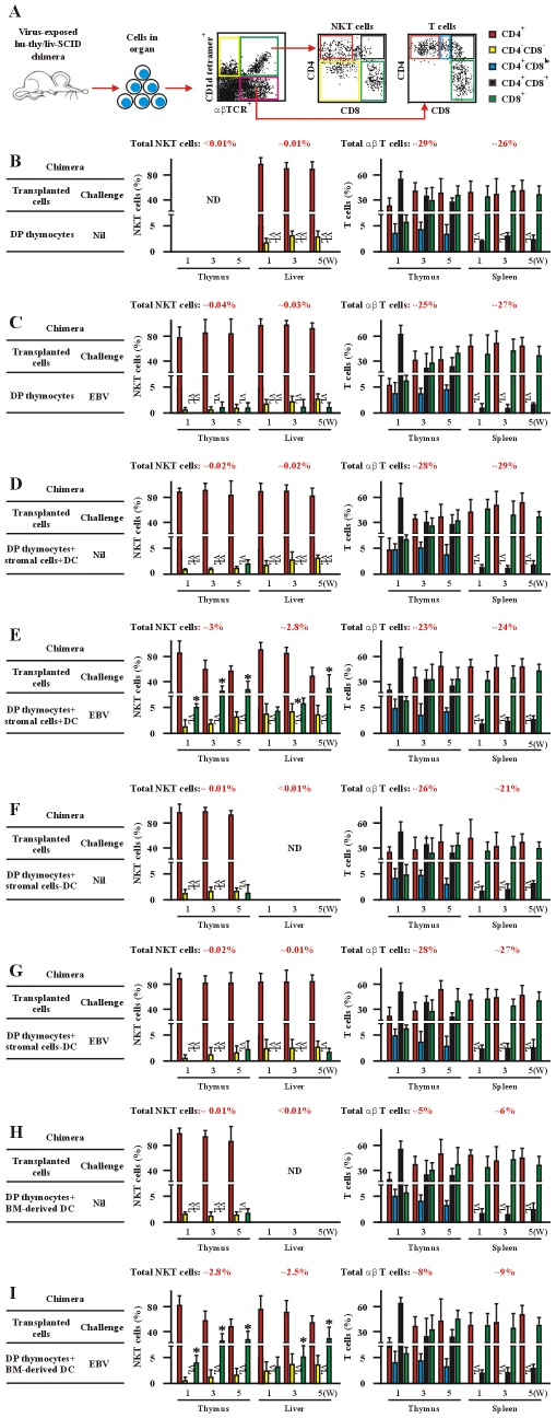
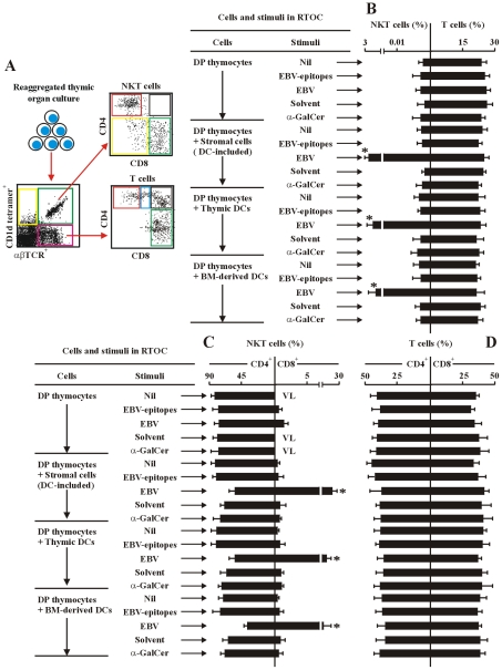
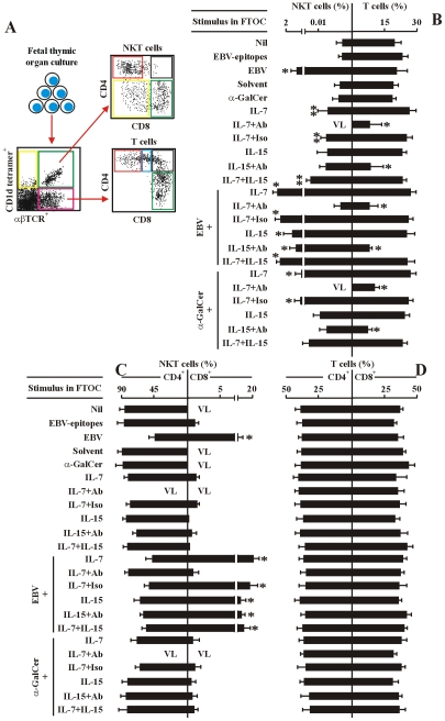
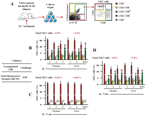
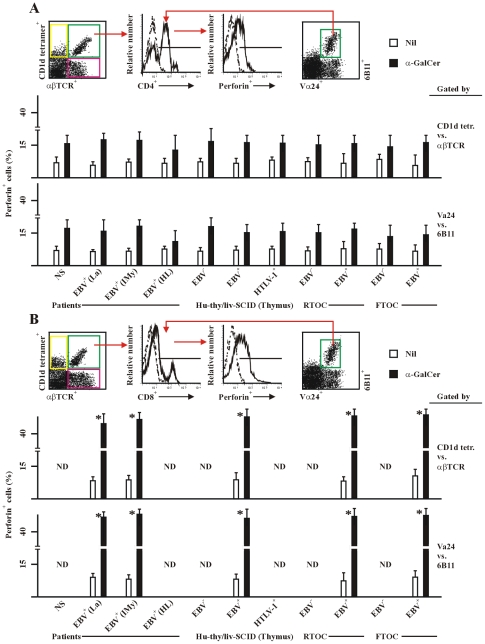
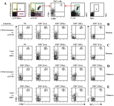
The reports on the origin of human CD8(+) Valpha24(+) T-cell receptor (TCR) natural killer T (NKT) cells are controversial. The underlying mechanism that controls human CD4 versus CD8 NKT cell development is not well-characterized. In the present study, we have studied total 177 eligible patients and subjects including 128 healthy latent Epstein-Barr-virus(EBV)-infected subjects, 17 newly-onset acute infectious mononucleosis patients, 16 newly-diagnosed EBV-associated Hodgkin lymphoma patients, and 16 EBV-negative normal control subjects. We have established human-thymus/liver-SCID chimera, reaggregated thymic organ culture, and fetal thymic organ culture. We here show that the average frequency of total and CD8(+) NKT cells in PBMCs from 128 healthy latent EBV-infected subjects is significantly higher than in 17 acute EBV infectious mononucleosis patients, 16 EBV-associated Hodgkin lymphoma patients, and 16 EBV-negative normal control subjects. However, the frequency of total and CD8(+) NKT cells is remarkably increased in the acute EBV infectious mononucleosis patients at year 1 post-onset. EBV-challenge promotes CD8(+) NKT cell development in the thymus of human-thymus/liver-SCID chimeras. The frequency of total (3% of thymic cells) and CD8(+) NKT cells ( approximately 25% of NKT cells) is significantly increased in EBV-challenged chimeras, compared to those in the unchallenged chimeras (<0.01% of thymic cells, CD8(+) NKT cells undetectable, respectively). The EBV-induced increase in thymic NKT cells is also reflected in the periphery, where there is an increase in total and CD8(+) NKT cells in liver and peripheral blood in EBV-challenged chimeras. EBV-induced thymic CD8(+) NKT cells display an activated memory phenotype (CD69(+)CD45RO(hi)CD161(+)CD62L(lo)). After EBV-challenge, a proportion of NKT precursors diverges from DP thymocytes, develops and differentiates into mature CD8(+) NKT cells in thymus in EBV-challenged human-thymus/liver-SCID chimeras or reaggregated thymic organ cultures. Thymic antigen-presenting EBV-infected dendritic cells are required for this process. IL-7, produced mainly by thymic dendritic cells, is a major and essential factor for CD8(+) NKT cell differentiation in EBV-challenged human-thymus/liver-SCID chimeras and fetal thymic organ cultures. Additionally, these EBV-induced CD8(+) NKT cells produce remarkably more perforin than that in counterpart CD4(+) NKT cells, and predominately express CD8alphaalpha homodimer in their co-receptor. Thus, upon interaction with certain viruses, CD8 lineage-specific NKT cells are developed, differentiated and matured intrathymically, a finding with potential therapeutic importance against viral infections and tumors.


Conflict of interest statement

TJ, HY, XR, XW, LL, and WL have a patent pending on a method to establish the EBV-challenged human-thymus/liver-SCID chimera for NKT cell research and therapeutic usage.

Figures

Figure 1
Figure 1. Human NKT and T cells in the various EBV-infected and non-infected subjects.
(A) The experimental and analysis scheme for detecting co-receptor-expressing NKT cells and T cells in PBMC was illustrated. The data to establish negative staining gates with fluorochrome conjugated empty CD1d tetramers (eCD1d tetramer) and αβTCR isotype mAb (αβTCRIso) control was shown in rightmost panel of this subfigure. (B) and (C) Frequencies of total (left panel) and co-receptor-expressing (right panel) NKT (B) and conventional αβT cells (C) in PBMCs from healthy latent EBV-infected subjects [EBV+(La)], newly-onset acute infectious mononucleosis patients [EBV+(IMa)], IM patients at year 1 post-onset [EBV+(IMy)], EBV-associated HL patients [EBV+(HL)] and EBV-negative normal control subjects (NS) assessed by flow cytometry. The absolute numbers per ml of NKT cell subsets of various patient and subject were shown in bottom panel as means ± s.d. (the s.d. was not shown for simplicity of the figure) (B). Data were mean ± s.d. (For patient numbers see Table S1). *, p<0.001.
Figure 2
Figure 2. EBV promotes CD8+ NKT cell development in vivo in hu-thy/liv-SCID chimeric mice.
(A) and (B) Development of total NKT (A) and T cells (B). The frequencies of NKT cells and T cells in thymus, liver, and spleen from hu-thy/liv-SCID chimeras challenged i.t. with EBV (EBV+) or HTLV-1 (HTLV-1+), were assessed by flow cytometry (middle and right panels) at the indicated timepoints. Unchallenged hu-thy/liv-SCID chimeras (Nil) were used as controls. Empty CD1d tetramer and isotype matched control Abs, which were used as staining controls, were not illustrated in this figure. The experimental and analysis scheme was shown in the leftmost panel. Data were mean ± s.d. (n  =  8). *, p<0.001. EBV-challenged chimeras vs. non-challenged or HTLV-1-challenged chimeras. (C) The experimental and analysis scheme for detecting the co-receptor-expressing NKT cells and T cells in various organs from different hu-thy/liv-SCID chimeras was illustrated. The α-GalCer-loaded CD1d tetramer, αβTCR mAb and other relevant mAbs were used to identify the different subsets of NKT cells and T cells as illustrated. The appropriate isotype Abs and empty fluorochrome-conjugated CD1d tetramer were used as controls. (D) and (E) Data showed the frequencies of co-receptor-expressing NKT cells and T cells in the unchallenged (D, Nil) or EBV challenged (E, EBV) hu-thy/liv-SCID chimeras. The protocols for the establishment of the chimeras were described in the leftmost panels. The chimeras were sacrificed at the indicated time intervals following post-immune-reconstitution and viral challenge and the various organs and tissues were collected. +DC, thymic DCs were included. VL, very low level (below detectable levels). Data were mean ± s.d. (n  =  7). *, p<0.001, EBV-challenged chimeras vs. non-challenged or HTLV-1-challenged chimeras.
Figure 3
Figure 3. EBV-induced CD8+ NKT cell development occurs at the DP thymocyte stage and depends upon thymic dendritic cells.
(A) The experimental and analysis scheme for detecting co-receptor-expressing NKT cells and T cells in various organs from different hu-thy/liv-SCID chimeras is illustrated after collecting 2.0×10⁶ total cell events. (BI) Development of co-receptor-expressing NKT cells (middle panels) and T cells (right panels). Data illustrate the frequency of co-receptor-expressing NKT cells and T cells in thymus, liver and spleen from EBV-challenged hu-thy/liv-SCID chimeras (EBV). Unchallenged hu-thy/liv-SCID chimeras (Nil) were used as controls. The protocols for the establishment of the various hu-thy/liv-SCID chimeras were described in the leftmost panels. (BG) Instead of total thymocytes, hu-thy/liv-SCID chimeras were established by intrathymic transplantation with DP thymocytes. The chimeras were sacrificed at the indicated time points following post-immune-reconstitution and viral challenge. Staining was performed as in Figure 2. +DC, thymic DCs were included. -DC, thymic DCs were depleted. In (H) and (I) the hu-thy/liv-SCID chimeras were established by intrathymic transplantation with DP thymocytes. Instead of transplantation with thymic stromal cells, BM-derived dendritic cells (1×10⁶ cells) were injected i.v. into the hu-thy/liv-SCID chimeras at t = 0. VL, very low level (below detectable level). ND, no determination. Data (BI) were mean ± s.d. (n  =  8). *, p<0.001, EBV-challenged chimeras vs. non-challenged chimeras.
Figure 4
Figure 4. EBV-induced CD8+ NKT cell differentiation depends upon thymic dendritic cells.
(A) The experimental and analysis scheme for detecting co-receptor-expressing NKT cells and T cells in various reaggregated fetal thymic organ cultures (RTOCs). After 14-days of culture, the various cell types were identified by flow cytometry as in Figure 2. (BD) Frequency of total NKT cells and total T cells (B), co-receptor-expressing NKT cells (C) and T cells (D), in the different RTOCs. The protocols for the establishment of the RTOCs were described in the leftmost panels in each sub-figure: DP thymocytes were reaggregated with either total thymic stromal cells (DC-included), purified thymic dendritic cells, or BM-derived dendritic cells. The stimuli were added as indicated. Nil, no stimulus; EBV-epitopes, HLA-A2-restricted, derived from the lytic cycle protein BMLF1 and HLA-DRB1-restricted, derived from nuclear antigen EBNA1 (10 µg/ml each); EBV, infectious EBV (107 pfu); Solvent, 0.005% polysorbate 20; α-GalCer (0.1 µg/ml). The RTOCs were harvested, and assessed by flow cytometry. Only the data for CD4+ and CD8+ NKT cells, and CD4 SP and CD8 SP T cells were shown. VL, very low level (below detectable level). Data were mean ± s.d. (n  =  10). *, p<0.001, EBV-challenged RTOCs vs. non-challenged RTOCs.
Figure 5
Figure 5. IL-7 is required for EBV-induced CD8+ NKT cell differentiation in vitro.
(A) The experimental and analysis scheme for detecting co-receptor-expressing NKT cells and T cells in various human fetal thymic organ cultures. After 14-day-culture, the various cell types were identified by flow cytometry as in Figure 2. (BD) Frequency of total NKT cells and total T cells (B), co-receptor-expressing NKT cells (C) and T cells (D) in the different FTOCs. The protocols for the establishment of different FTOCs were described in the leftmost panels in each sub-figure. The various stimuli and blockers were added as indicated. Nil, no stimulus; EBV-epitopes, HLA-A2-restricted, derived from the lytic cycle protein BMLF1 and HLA-DRB1-restricted, derived from nuclear antigen EBNA1 (1 µg/ml each); EBV, infectious EBV (107 pfu); Solvent, 0.005% polysorbate 20; α-GalCer (0.1 µg/ml). IL-7 (10 ng/ml); IL-15 (10 ng/ml), Abs, either mouse anti-human IL-7 monoclonal Ab plus mouse anti-human IL-7Rα monoclonal Ab, or mouse anti-human IL-15 monoclonal Ab plus mouse anti-human IL-15Rα monoclonal Ab, respectively (5 µg/ml each); The FTOCs were harvested and assessed by flow cytometry. Only the data for CD4+ and CD8+ NKT cells and CD4 SP and CD8 SP T cells were shown. VL, very low level (below detectable level). Data were mean ± s.d. (n = 10). ** p<0.05. *, p<0.001, EBV-challenged FTOCs vs. non-challenged FTOCs; IL-7-challenged FTOCs vs. non-challenged FTOCs.
Figure 6
Figure 6. IL-7 is required for EBV-induced CD8+ NKT cell development in vivo.
(A) The experimental and analysis scheme for detecting co-receptor-expressing NKT cells and T cells in various organs from hu-thy/liv-SCID chimeras treated with IL-7. The chimeras were established by intrathymic transplantation with DP thymocytes. The chimeras were treated i.v. with IL-7 (0.1 µg/kg/d), or with IL-7 plus mAb against IL-7 (1 µg/kg/d) (IL-7+Ab), or IL-7 plus isotype Ab (1 µg/kg/d) (IL-7+Iso) as indicated. The chimeras were sacrificed at the indicated time points following post-immune-reconstitution and viral challenge. Staining was performed as in Figure 2. (BD) Development of co-receptor-expressing NKT cells. Data show the frequency of co-receptor-expressing NKT cells in thymus or livers from hu-thy/liv-SCID chimera challenged i.t. with EBV (EBV), as assessed by flow cytometry. The protocols for the establishment of the chimeras were described in the leftmost panel. +DC, thymic DCs were included. VL, very low level (below detectable level). Data (BD) were mean ± s.d. (n = 7 or 8). *, p<0.001, EBV- and IL-7-challenged chimeras vs. a-IL-7 Ab-treated chimeras.
Figure 7
Figure 7. Perforin expression in human and chimeric CD4+ and CD8+ NKT cells.
PBMC were from healthy latent EBV-infected subjects [EBV+(La)], IM patients at year 1 post-onset [EBV+(IMy)], EBV-associated HL patients [EBV+(HL)] and EBV-negative normal control subjects (NS). Thymic cell suspension were from EBV-exposed (EBV+), un-exposed (EBV) or HTLV-1-exposed (HTLV-1+) hu-thy/liv-SCID chimeras, or from EBV-exposed (EBV+) or un-exposed (EBV) RTOC and FTOC. For detection of intracellular expression of perforin, cells were stimulated with α-GalCer (1 µg/ml) for 24 hrs, intracellular stained, and assessed by flow cytometry using the experimental strategy shown in the upper panel of each subfigure. The NKT cells were gated by either CD1d tetramers vs. anti-αβTCR mAb (middel panels) or anti-Vα24 mAb vs. 6B11 mAb (bottom panels). Perforin positive cells (%) in gated CD4+ (A) and CD8+ (B) NKT cells were shon. Solvent for α-GalCer was 0.005% polysorbate 20 (not shown). Nil, negative stimulation control. ND, no determination. Data were mean ± s.d. (n = 8). **, p<0.05; *, p<0.001. CD8+ in (B) vs. counterpart CD4+ NKT cells in (A).
Figure 8
Figure 8. NKT cells differentially express the CD8α and CD8β chain.
(A) The experimental and analysis scheme for detecting total and co-receptor CD8α- and CD8β-expressing NKT cells. (B) and (C) NKT cells in PBMCs from healthy latent EBV-infected subjects [EBV+(La)], IM patients at year 1 post-onset [EBV+(IMy)], EBV-associated HL patients [EBV+(HL)] and EBV-negative normal control subjects (NS) were assessed by flow cytometry using the gate of either CD1d tetramers vs. anti-αβTCR mAb (B) or anti-Vα24 mAb vs. 6B11 mAb (C). Further dot plot analysis of CD8α vs. CD8β in gated NKT cells was shown. (D) and (E), NKT cells in thymus (Thy) and liver (Liv) from hu-thy/liv-SCID chimeras challenged i.t. with EBV (EBV+) or unchallenged (EBV) were assessed by flow cytometry using the gate of either CD1d tetramers vs. anti-αβTCR mAb (D) or anti-Vα24 mAb vs. 6B11 mAb (E). Further dot plot analysis of CD8α vs. CD8β in gated NKT cells was shown. Data were representatives of 5 similar experiments in each group.

Similar articles

Cited by

References

    1. Kronenberg M. Toward an understanding of NKT cell biology: progress and paradoxes. Annu Rev Immunol. 2005;23:877–900. - PubMed
    1. Bendelac A, Savage PB, Teyton L. The biology of NKT cells. Annu Rev Immunol. 2007;25:297–336. - PubMed
    1. Godfrey DI, Kronenberg M. Going both ways: immune regulation via CD1d-dependent NKT cells. J Clin Invest. 2004;114:1379–1388. - PMC - PubMed
    1. Brigl M, Brenner MB. CD1: antigen presentation and T cell function. Annu Rev Immunol. 2004;22:817–890. - PubMed
    1. Zhou D, Mattner J, Cantu C, 3rd, Schrantz N, Yin N, et al. Lysosomal glycosphingolipid recognition by NKT cells. Science. 2004;306:1786–1789. - PubMed

Publication types

MeSH terms