

Skeletal site-related variation in human trabecular bone transcriptome and signaling
- PMID: 20502692
- PMCID: PMC2872667
- DOI: 10.1371/journal.pone.0010692
Skeletal site-related variation in human trabecular bone transcriptome and signaling
Abstract
Background: The skeletal site-specific influence of multiple genes on bone morphology is recognised, but the question as to how these influences may be exerted at the molecular and cellular level has not been explored.
Methodology: To address this question, we have compared global gene expression profiles of human trabecular bone from two different skeletal sites that experience vastly different degrees of mechanical loading, namely biopsies from iliac crest and lumbar spinal lamina.
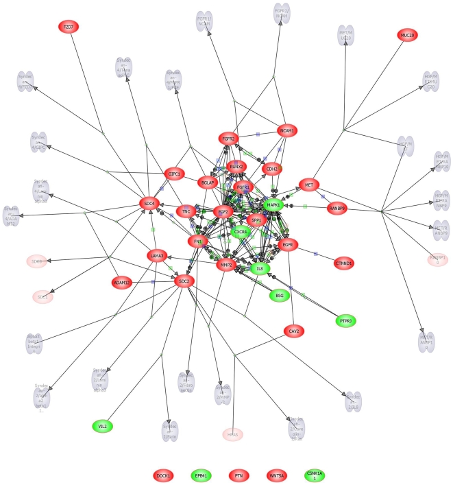
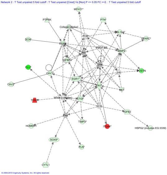
Principal findings: In the lumbar spine, compared to the iliac crest, the majority of the differentially expressed genes showed significantly increased levels of expression; 3406 transcripts were up- whilst 838 were down-regulated. Interestingly, all gene transcripts that have been recently demonstrated to be markers of osteocyte, as well as osteoblast and osteoclast-related genes, were markedly up-regulated in the spine. The transcriptome data is consistent with osteocyte numbers being almost identical at the two anatomical sites, but suggesting a relatively low osteocyte functional activity in the iliac crest. Similarly, osteoblast and osteoclast expression data suggested similar numbers of the cells, but presented with higher activity in the spine than iliac crest. This analysis has also led to the identification of expression of a number of transcripts, previously known and novel, which to our knowledge have never earlier been associated with bone growth and remodelling.
Conclusions and significance: This study provides molecular evidence explaining anatomical and micro-architectural site-related changes in bone cell function, which is predominantly attributable to alteration in cell transcriptional activity. A number of novel signaling molecules in critical pathways, which have been hitherto not known to be expressed in bone cells of mature vertebrates, were identified.
Conflict of interest statement
Figures


Similar articles
-
Osteocyte-Mediated Translation of Mechanical Stimuli to Cellular Signaling and Its Role in Bone and Non-bone-Related Clinical Complications.Curr Osteoporos Rep. 2020 Feb;18(1):67-80. doi: 10.1007/s11914-020-00564-9. Curr Osteoporos Rep. 2020. PMID: 31953640 Review.
-
Zic1 transcription factor in bone: neural developmental protein regulates mechanotransduction in osteocytes.FASEB J. 2010 Aug;24(8):2893-903. doi: 10.1096/fj.09-148908. Epub 2010 Mar 30. FASEB J. 2010. PMID: 20354137
-
Effects of Age and Estrogen on Skeletal Gene Expression in Humans as Assessed by RNA Sequencing.PLoS One. 2015 Sep 24;10(9):e0138347. doi: 10.1371/journal.pone.0138347. eCollection 2015. PLoS One. 2015. PMID: 26402159 Free PMC article.
-
Human primary osteocyte differentiation in a 3D culture system.J Bone Miner Res. 2009 Nov;24(11):1927-35. doi: 10.1359/jbmr.090517. J Bone Miner Res. 2009. PMID: 19419324
-
The Osteocyte Transcriptome: Discovering Messages Buried Within Bone.Curr Osteoporos Rep. 2021 Dec;19(6):604-615. doi: 10.1007/s11914-021-00708-5. Epub 2021 Nov 10. Curr Osteoporos Rep. 2021. PMID: 34757588 Free PMC article. Review.
Cited by
-
Bone Anabolic Response in the Calvaria Following Mild Traumatic Brain Injury is Mediated by the Cannabinoid-1 Receptor.Sci Rep. 2019 Nov 7;9(1):16196. doi: 10.1038/s41598-019-51720-w. Sci Rep. 2019. PMID: 31700010 Free PMC article.
-
Genetic Polymorphism of miR-196a-2 is Associated with Bone Mineral Density (BMD).Int J Mol Sci. 2017 Nov 25;18(12):2529. doi: 10.3390/ijms18122529. Int J Mol Sci. 2017. PMID: 29186852 Free PMC article.
-
Mechanical-Stress-Related Epigenetic Regulation of ZIC1 Transcription Factor in the Etiology of Postmenopausal Osteoporosis.Int J Mol Sci. 2022 Mar 9;23(6):2957. doi: 10.3390/ijms23062957. Int J Mol Sci. 2022. PMID: 35328378 Free PMC article.
-
The Influence of DNA Methylation on Bone Cells.Curr Genomics. 2015 Dec;16(6):384-92. doi: 10.2174/1389202916666150817202913. Curr Genomics. 2015. PMID: 27019613 Free PMC article.
-
Human osteoblasts obtained from distinct periarticular sites demonstrate differences in biological function in vitro.Bone Joint Res. 2021 Sep;10(9):611-618. doi: 10.1302/2046-3758.109.BJR-2020-0497.R1. Bone Joint Res. 2021. PMID: 34565180 Free PMC article.
References
-
- Lanyon LE. Functional strain as a determinant of bone remodeling. Calcif Tissue Int. 1984;36:S56–S61. - PubMed
-
- Rubin CT, Lanyon LE. Regulation of bone mass by mechanical strain magnitude. Calcif Tissue Int. 1985;37:411–417. - PubMed
-
- Mannion AF, Adams MA, Dolan P. Sudden and unexpected loading generates high forces on the LS. Spine. 2000;25:842–852. - PubMed
-
- Turner CH, Pavalko FM. Mechanotransduction and functional response of the skeleton to physical stress: the mechanisms and mechanics of bone adaptation. J Orthop Sci. 1998;3:346–355. - PubMed
-
- Huiskes R, Ruimerman R, van Lenthe GH, Janssen JD. Effects of mechanical forces on maintenance and adaptation of form in trabecular bone. Nature. 2000;405:704–706. - PubMed
Publication types
MeSH terms
Substances
LinkOut - more resources
Full Text Sources
