

Nerve sheath myxoma (neurothekeoma) of the gingiva, a case report and review of the literature
- PMID: 20502996
- PMCID: PMC2923317
- DOI: 10.1007/s12105-010-0183-5
Nerve sheath myxoma (neurothekeoma) of the gingiva, a case report and review of the literature
Abstract
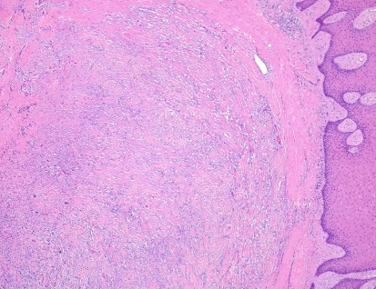
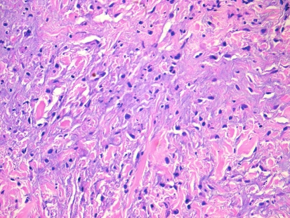
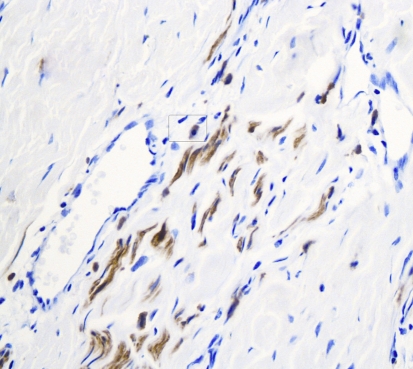
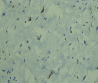
Nerve sheath myxoma (NSM) is a benign peripheral nerve sheath tumor that rarely occurs in the oral cavity. Among the 23 reported intraoral cases, no lesion has previously been reported on the gingiva. In this report, we describe the first gingival case of oral neurothekeoma with histopathologic and immunohistochemical characteristics. The patient, a 32 year old female presented with a slowly growing gingival mass diagnosed clinically as an epulis. The lesion was surgically excised. Histopathologically, the lesion presented as a submucosal multinodular mass composed of spindle and stellate-shaped cells with a myxoid background. Immunohistochemically, the tumor cells were sporadically positive for S-100 and NSE and negative for GFAP, EMA, SMA, CD68 and HMB45. The immunoprofile of this lesion confirmed a Schwann cell origin. The lesion was followed up for 10 months with no reports of recurrence.
Figures
References
-
- Harkin JC, Reed JJ. Tumors of the peripheral nervous system, second series, Fasc, 3. Washington, D. C: Armed Forces Institute of Pathology; 1969. pp. 60–64.
-
- Gallagher RL, Helwig EB. Neurothekeoma-a benign cutaneous tumor of neural origin. Am J Clin Pathol. 1980;74:759–761. - PubMed
Publication types
MeSH terms
Substances
LinkOut - more resources
Full Text Sources
Research Materials
Miscellaneous
