

Characterization of CD4+ T cells specific for glutamic acid decarboxylase (GAD65) and proinsulin in a patient with stiff-person syndrome but without type 1 diabetes
- PMID: 20503259
- PMCID: PMC2878280
- DOI: 10.1002/dmrr.1083
Characterization of CD4+ T cells specific for glutamic acid decarboxylase (GAD65) and proinsulin in a patient with stiff-person syndrome but without type 1 diabetes
Abstract
Background: Glutamic acid decarboxylase (GAD) is a rate-limiting enzyme in the synthesis of gamma-amino butyric acid (GABA) and an important autoantigen both in patients with type 1 diabetes (T1D) and stiff-person syndrome (SPS). Autoantibodies (GADA) to the 65-kDa isoform of GAD are a characteristic feature in both diseases. Approximately 30% of patients with SPS develop diabetes, yet, it is unclear to which extent co-existing autoimmunity to GAD65 and other islet autoantigens determines the risk of developing T1D.
Methods: In this study, we monitored CD4+ T-cell responses to GAD65 and proinsulin in a patient with SPS who remained normoglycaemic during the 46-month follow-up.
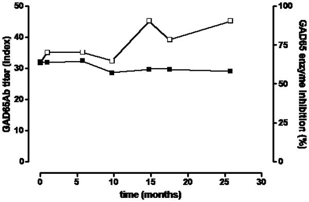
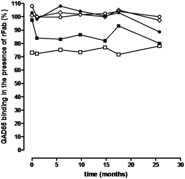
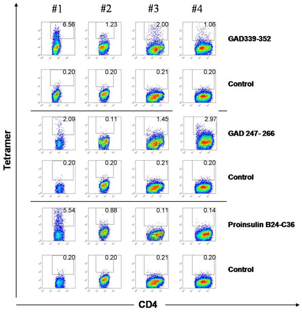
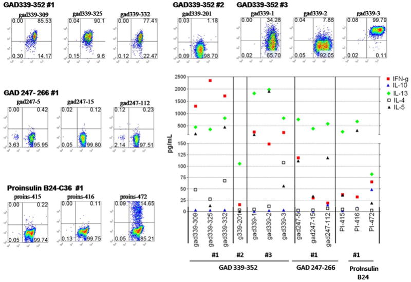
Results: Fluctuating but persistent T-cell reactivity to GAD65 was identified, as well as T-cell reactivity to proinsulin at one time point. The majority of the T-cell clones isolated from the patient with SPS produced high levels of Th2 cytokines (IL-13, IL-5 and IL-4). We also examined levels of GADA, insulin and IA-2 autoantibodies, and epitope specificity of GADA. In both serum and cerebrospinal fluid (CSF), GADA levels were high, and GADA persisted throughout the follow-up. Despite T-cell reactivity to both GAD65 and proinsulin, autoantibodies to other islet autoantigens did not develop.
Conclusions: Further follow-up will determine whether the beta-cell autoimmunity observed in this patient will eventually lead to T1D.
Figures
Similar articles
-
Higher autoantibody levels and recognition of a linear NH2-terminal epitope in the autoantigen GAD65, distinguish stiff-man syndrome from insulin-dependent diabetes mellitus.J Exp Med. 1994 Aug 1;180(2):595-606. doi: 10.1084/jem.180.2.595. J Exp Med. 1994. PMID: 7519242 Free PMC article.
-
Characteristics of in-vitro phenotypes of glutamic acid decarboxylase 65 autoantibodies in high-titre individuals.Clin Exp Immunol. 2013 Mar;171(3):247-54. doi: 10.1111/cei.12026. Clin Exp Immunol. 2013. PMID: 23379430 Free PMC article. Clinical Trial.
-
Stiff-person syndrome (SPS) and anti-GAD-related CNS degenerations: protean additions to the autoimmune central neuropathies.J Autoimmun. 2011 Sep;37(2):79-87. doi: 10.1016/j.jaut.2011.05.005. Epub 2011 Jun 16. J Autoimmun. 2011. PMID: 21680149 Review.
-
GAD65 IgG autoantibodies in stiff person syndrome: clonality, avidity and persistence.Eur J Neurol. 2008 Sep;15(9):973-80. doi: 10.1111/j.1468-1331.2008.02221.x. Epub 2008 Jul 10. Eur J Neurol. 2008. PMID: 18637036
-
Immunobiology of stiff-person syndrome.Int Rev Immunol. 2008 Jan-Apr;27(1-2):79-92. doi: 10.1080/08830180701883240. Int Rev Immunol. 2008. PMID: 18300057 Review.
Cited by
-
The limbic and extra-limbic encephalitis associated with glutamic acid decarboxylase (GAD)-65 antibodies: an observational study.Neurol Sci. 2025 Jun;46(6):2765-2777. doi: 10.1007/s10072-024-07933-7. Epub 2024 Dec 20. Neurol Sci. 2025. PMID: 39704979
-
Mechanistic basis of immunotherapies for type 1 diabetes mellitus.Transl Res. 2013 Apr;161(4):217-29. doi: 10.1016/j.trsl.2012.12.017. Epub 2013 Jan 22. Transl Res. 2013. PMID: 23348026 Free PMC article. Review.
-
Production of primary human CD4⁺ T cell lines and clones.Methods Mol Biol. 2013;960:545-555. doi: 10.1007/978-1-62703-218-6_40. Methods Mol Biol. 2013. PMID: 23329513 Free PMC article.
-
Autoimmune polyglandular syndrome and GAD65 related-temporal lobe epilepsy.Neurol Sci. 2025 Jun 26. doi: 10.1007/s10072-025-08330-4. Online ahead of print. Neurol Sci. 2025. PMID: 40571861
-
Autoimmune stiff person syndrome and related myelopathies: understanding of electrophysiological and immunological processes.Muscle Nerve. 2012 May;45(5):623-34. doi: 10.1002/mus.23234. Muscle Nerve. 2012. PMID: 22499087 Free PMC article. Review.
References
-
- Duddy ME, Baker MR. Stiff person syndrome. Front Neurol Neurosci. 2009;26:147–65. - PubMed
-
- Knip M, Siljander H. Autoimmune mechanisms in type 1 diabetes. Autoimmun Rev. 2008;7 :550–7. - PubMed
-
- Skyler JS. Diabetes mellitus: pathogenesis and treatment strategies. J Med Chem. 2004;47 :4113–7. - PubMed
-
- Dinkel K, Meinck HM, Jury KM, Karges W, Richter W. Inhibition of gamma-aminobutyric acid synthesis by glutamic acid decarboxylase autoantibodies in stiff-man syndrome. Ann Neurol. 1998;44:194–201. - PubMed
Publication types
MeSH terms
Substances
Grants and funding
LinkOut - more resources
Full Text Sources
Other Literature Sources
Research Materials
