. 2010 May;26(4):306-18.
doi: 10.1002/dmrr.1088.

Diet-induced obesity in Sprague-Dawley rats causes microvascular and neural dysfunction

Affiliations

Diet-induced obesity in Sprague-Dawley rats causes microvascular and neural dysfunction

Eric P Davidson et al. Diabetes Metab Res Rev. 2010 May.

Abstract

Background: The objective of this study was to determine the effect of diet-induced obesity (DIO) on microvascular and neural function.

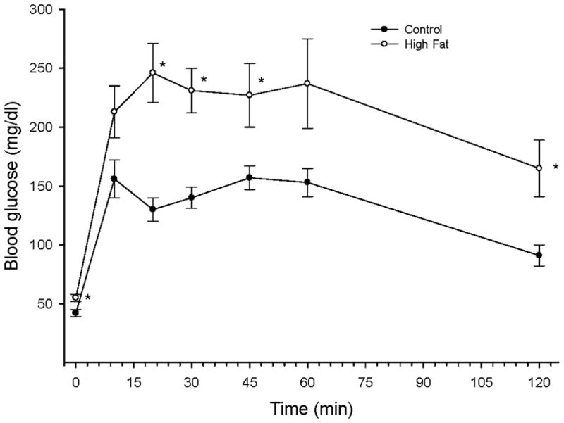
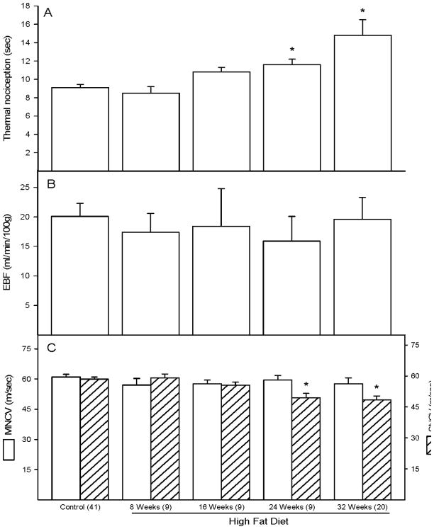
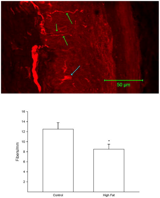
Methods: Rats were fed a standard or high fat diet for up to 32 weeks. The following measurements were carried out: vasodilation in epineurial arterioles using videomicroscopy, endoneurial blood flow using hydrogen clearance, nerve conduction velocity using electrical stimulation, size-frequency distribution of myelinated fibres of the sciatic nerve, intraepidermal nerve fibre density using confocal microscopy and thermal nociception using the Hargreaves method.

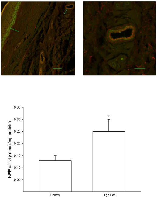
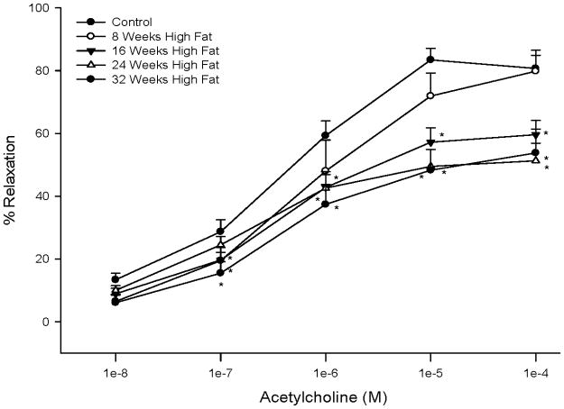
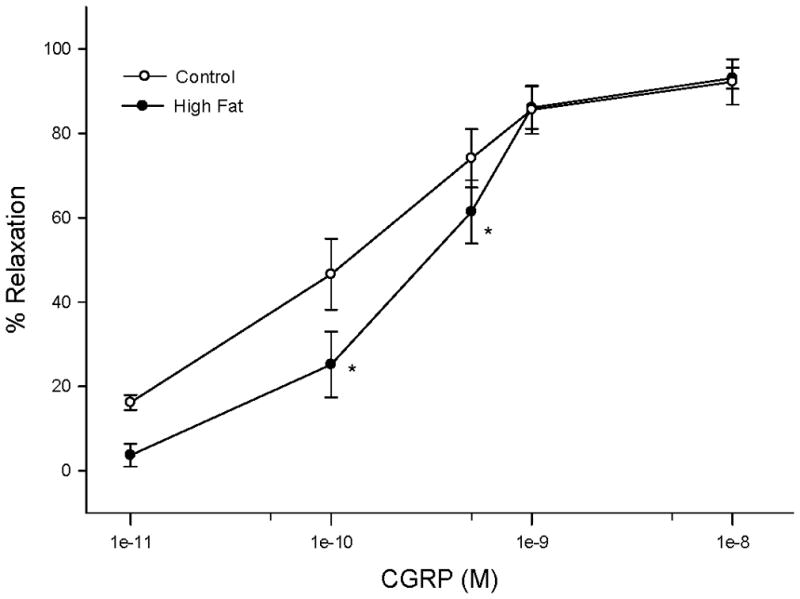
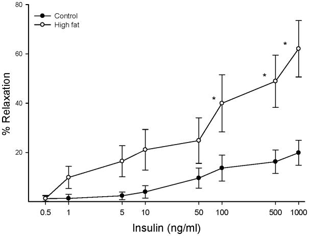
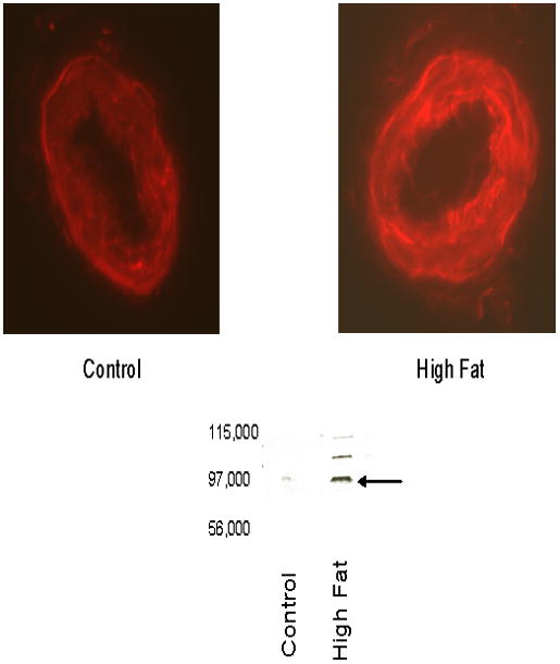
Results: Rats fed a high fat diet for 32 weeks developed sensory neuropathy, as indicated by slowing of sensory nerve conduction velocity and thermal hypoalgesia. Motor nerve conduction velocity and endoneurial blood flow were not impaired. Mean axonal diameter of myelinated fibres of the sciatic nerve was unchanged in high fat-fed rats compared with that in control. Intraepidermal nerve fibre density was significantly reduced in high fat-fed rats. Vascular relaxation to acetylcholine and calcitonin gene-related peptide was decreased and expression of neutral endopeptidase (NEP) increased in epineurial arterioles of rats fed a high fat diet. In contrast, insulin-mediated vascular relaxation was increased in epineurial arterioles. NEP activity was significantly increased in the skin of the hindpaw. Markers of oxidative stress were increased in the aorta and serum of high fat-fed rats but not in epineurial arterioles.

Conclusion: Chronic obesity causes microvascular and neural dysfunction. This is associated with increased expression of NEP but not oxidative stress in epineurial arterioles. NEP degrades vasoactive peptides, which may explain the decrease in microvascular function.


Figures

Figure 1
Figure 1. Effect of a high fat diet on glucose tolerance
Rats were fed a standard or high fat diet for 32 weeks. Afterwards glucose tolerance was determined as described in the Methods section. Data are presented as the mean ± SEM in mg/dl. * p < 0.05, for individual data points compared to rats fed a standard diet (control). The area under the curve (AUC) was significantly different p < 0.01 for high fat fed rats vs. control. The number of rats in each group was the same as shown in Table 1.
Figure 2
Figure 2. Effect of a high fat diet on thermal nociception, endoneurial blood flow and motor and sensory nerve conduction velocity
Data are presented as the mean ± SEM for thermal nociception in sec (A), nutritive blood flow in ml/min/100g (B), and motor and sensory nerve conduction velocity in m/sec (C). The number of experimental determinations is presented in parentheses. For these studies control rats were age matched for each time point. Data from each of these time points for the control rats were not affected by age so these data were combined. * p < 0.05, compared to rats fed the standard diet (control).
Figure 3
Figure 3. Effect of a high fat diet on intraepidermal nerve fiber profiles
Rats were fed a standard or high fat diet for 32 weeks. Afterwards, the number of intraepidermal nerve fiber profiles were determined as described in the Methods section. A representative image for intraepidermal nerve fiber profiles is provided (top). The three green arrows point at individual IENF profiles. The blue arrow near the bottom of the image points at a Langerhans cell. The number of rats in each group was the same as shown in Table 1. * p < 0.05, compared to rats fed the standard diet (control).
Figure 4
Figure 4. Effect of a high fat diet on neutral endopeptidase activity in the skin of the hindpaw
Rats were fed a standard or high fat diet for 32 weeks. Afterwards, activity of neutral endopeptidase in a skin biopsy of the hindpaw was determined as described in the Methods section. A representative image for expression of neutral endopeptidase in the hindpaw is provided (top). The left image (inserted bar 50 μm) demonstrates the staining for neutral endopeptidase (green) that occurs in basal keratinocytes in the stratum basale (green arrow on left). The blue arrow pointing at the vessel in the upper right portion of the image on the left side is enlarged and shown on the right side. The right image (inserted bar 20 μm) demonstrates that staining for VWF occurring in the endothelium (red staining) does not overlap with the staining for neutral endopeptidase (green staining) suggesting that neutral endopeptidase in the vasculature is primarily located in the smooth muscle layer. The number of rats in each group was the same as shown in Table 1. * p < 0.05, compared to rats fed the standard diet (control).
Figure 5
Figure 5. Effect of a high fat diet on acetylcholine-mediated vascular relaxation of epineurial arterioles
Pressurized arterioles (40 mm Hg and ranging from 60–100 μm luminal diameter) were constricted with U46619 (30–50%) and incremental doses of acetylcholine were added to the bathing solution while recording steady state vessel diameter. Data are presented as the mean of % relaxation ± SEM. For these studies two vessels were collected from each rat, studied and the data combined. * p < 0.05, compared to rats fed the standard diet (control). The minimum number of rats in each group is 9.
Figure 6
Figure 6. Effect of a high fat diet on calcitonin gene-related peptide (CGRP) mediated vascular relaxation of epineurial arterioles
Arterioles were derived from control 32 week high fat fed rats as described in Figure 5. Incremental doses of CGRP were added to the bathing solution while recording steady state vessel diameter. Data are presented as the mean of % relaxation ± SEM. The number of rats in each group was the same as shown in Table 1. * p < 0.05, compared to rats fed the standard diet (control).
Figure 7
Figure 7. Effect of a high fat diet on insulin-mediated vascular relaxation by epineurial arterioles
Arterioles were derived from control 32 week high fat fed rats as described in Figure 5. Incremental doses of insulin were added to the bathing solution while recording steady state vessel diameter. The number of rats in each group was the same as shown in Table 1. Data are presented as the mean of % relaxation ± SEM. * p < 0.05, compared to rats fed the standard diet (control).
Figure 8
Figure 8. Effect of a high fat diet on expression of neutral endopeptidase in epineurial arterioles
Presented are representative fluorescent photomicrographs of confocal microscopic sections of epineurial arterioles of the sciatic nerve for neutral endopeptidase immunostaining (top) and Western blot analysis (bottom). The arrow points to the NEP band in the Western blot. The identity of the two other bands in the Western blot from the high fat fed sample is unknown but may be higher molecular weight isoforms. Each of the two vessels was examined on the same day using identical laser and photomultiplier settings. The experiment was repeated three times and the fold increase in expression of NEP in epineurial arterioles from rats fed a high fat diet was 2.6 ± 0.6 as determined by analysis of the immunostaining.

Similar articles

Cited by

References

    1. Caglayan E, Blaschke F, Takata Y, Hsueh WA. Metabolic syndrome-interdependence of the cardiovascular and metabolic pathways. Curr Opin Pharmacol. 2005;5:135–142. - PubMed
    1. Costa LA, Canani LH, Lisboa HR, Tres GS, Gross JL. Aggregation of features of the metabolic syndrome is associated with increased prevalence of chronic complications in Type 2 diabetes. Diabetic Med. 2004;21:252–255. - PubMed
    1. de Jongh RT, Serne EH, IJzerman RG, de Vries G, Stehouwer SD. Impaired microvascular function in obesity: implications for obesity-associated microangiopathy, hypertension, and insulin resistance. Circulation. 2004;109:2529–2535. - PubMed
    1. Oltman CL, Coppey LJ, Gellett JS, Davidson EP, Lund DD, Yorek MA. Progression of vascular and neural dysfunction in sciatic nerves of Zucker Diabetic Fatty (ZDF) and Zucker rats. Am J Physiol. 2005;289:E113–122. - PubMed
    1. LeRoith D, Rayfield EJ. The benefits of tight glycemic control in type 2 diabetic mellitus. Clinical Cornerstone. 2007;8:S19–S29. - PubMed

Publication types