. 2010 Jun;293(6):938-46.
doi: 10.1002/ar.20805.

Temporal changes in expression of FoxA1 and Wnt7A in isolated adult human alveolar epithelial cells enhanced by heparin

Affiliations

Temporal changes in expression of FoxA1 and Wnt7A in isolated adult human alveolar epithelial cells enhanced by heparin

K B C Apparao et al. Anat Rec (Hoboken). 2010 Jun.

Abstract

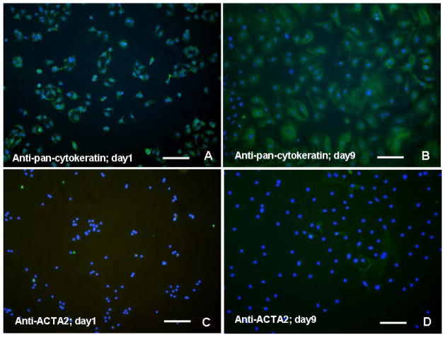
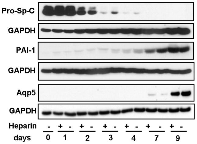
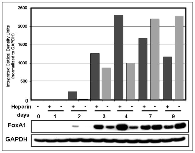
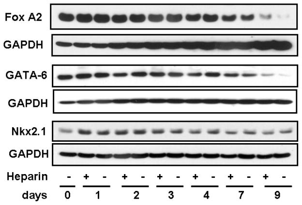
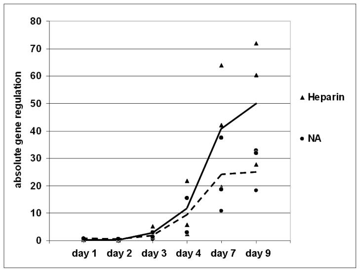
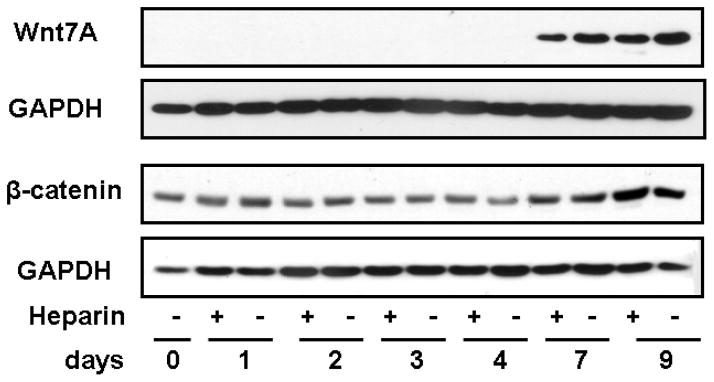
Pre- and postnatal developmental studies of the lung have provided compelling evidence demonstrating multiple factors that orchestrate alveolar epithelial cell differentiation. The extent to which reactivation of certain developmental pathways in the adult might influence the course of differentiation of alveolar type 2 cells (AT2) into AT1 cells is not known. In this study, we examined selected members of the forkhead (Fox) family of transcription factors and the Wnt (wingless) family of signaling proteins for expression during human alveolar cell differentiation in vitro and determined their potential responses to sulfated components of extracellular matrix (ECM), like those shed from cell surfaces or found in basement membrane and modeled by heparin. Isolated adult human AT2 cells cultured over a 9-day period were used to define the temporal profile of expression of targeted factors during spontaneous differentiation to AT1-like cells. FoxA1 protein was upregulated at early to intermediate time points, where it was strongly elevated by heparin. Gene expression of wnt7A increased dramatically beginning on day 3 and was enhanced even further on days 7 and 9 by heparin, whereas protein expression appeared at days 7 and 9. These temporal changes of expression suggest that sulfated ECMs may act to enhance the increase in FoxA1 at the critical juncture when AT2 cells commence the differentiation process to AT1 cells, in addition to enhancing the increase in wnt7A when the AT1 cell phenotype stabilizes. Collectively, these factors may act to modulate differentiation in the adult human pulmonary alveolus.


Figures

Figure 1
Figure 1
Immunofluorescent detection of pan-cytokeratin and smooth muscle actin (ACTA2) in isolated human alveolar cell preparations counterstained with DAPI. (A) Over 95% of “day 1” cells appear to be pan-cytokeratin positive, reflective of their epithelial origin; (B) Over 90% of “day 9” cells remained pan-cytokeratin positive, although significantly larger in size than “day 1” cells. (C) Cells positive for the myofibroblast marker ACTA2 at “day 1” in culture are difficult to resolve; (D) Only a few scattered myofibroblasts staining for ACTA2 in “day 9” cells confirm the relative purity of the AT2 isolation. Bar in each = 80 μm.
FIGURE 2
FIGURE 2
Time course of markers of alveolar epithelium differentiation from AT2 to AT1 cells. Prosurfactant protein-C (pro-SP-C; marker for AT2 cells), is highly expressed at days 0 and 1, but shows a distinct reduction beginning on day 2, although slightly less with heparin. At days 3 and 4, heparin treatment accelerates the increasing reduction of pro-SP-C. No pro-SP-C expression is evident at days 7 and 9. Plasminogen activator inhibitor-1 (PAI-1; marker for AT1 cells), was faintly detectable at day 1 and steadily increased as the time course progressed to day 9. Another marker for AT1 cells is aquaporin5 (Aqp5), which is detectable at day 7, but was very evident by day 9. GAPDH of the same blots is shown for normalization. Blots shown are representative of the similar results of three separate experiments.
FIGURE 3
FIGURE 3
FoxA1 protein expression is affected by heparin. FoxA1 protein in whole cell lysates of isolated human AT2 cells was analyzed by Western blot. Levels of FoxA1 steadily increased from day 2 in untreated cells, as confirmed in the densitometric tracings (gray bars). FoxA1 was highly increased by heparin treatment (black bars) at days 2, 3, and 4 but was decreased on days 7 and 9 by heparin. GAPDH of the same blot is shown as a loading control. For normalization of FoxA1 densitometry, equal areas of GAPDH signal in each lane were quantified to avoid signal overlap.
FIGURE 4
FIGURE 4
Other differentiation-related transcription factors in human alveolar cells are less affected by heparin. FoxA2 protein expression peaked early in the time course, and steadily decreased after day 4 and was relatively insensitive to heparin. GATA-6 protein expression peaked early in the time course and diminished through day 9. TTF-1 expression was low at isolation but strongly increased on day 1 postisolation, was moderately enhanced only at this early time point by heparin, and then tapered off toward day 9. Blots shown are representative of three similar results.
FIGURE 5
FIGURE 5
Wnt7A expression in isolated human alveolar type 2 cells varies with time and heparin treatments. Quantitative real time polymerase chain reaction (qRT-PCR) using TaqMan primers and probes was performed on cDNA equivalent to 100 ng of total RNA isolated from human AT2 cells cultured with or without 500 μg/mL heparin for 0–9 days. The absolute wnt7A gene regulation in cells from three individuals treated in separate experiments with (▲) or without (●) heparin is depicted at each time point. The solid line (—) connects the means of the expression at each time point in heparin-treated cells from the three individuals while the broken line (---) connects the means of expression at each time point in untreated cells. Wnt7A clearly increased rapidly from day 2 through day 9 and was enhanced by heparin on days 7 and 9.
FIGURE 6
FIGURE 6
Wnt7A and β-catenin protein expression in whole cell lysates of isolated human alveolar cells, analyzed by Western blot. Wnt7A protein expression was not detected by Western blot until day 7 and increased on day 9. β-catenin protein expression remained relatively steady from day 0 through day 4 and then was elevated at days 7 and 9, confirming activation of Wnt signaling. Each of these proteins is shown with the GAPDH band from the same blot for normalization; the temporal pattern was the same in each of three individuals, and representative blots are shown.

References

    1. Baeg GH, Lin X, Khare N, Baumgartner S, Perrimon N. Heparan sulfate proteoglycans are critical for the organization of the extracellular distribution of wingless. Development. 2001;128:87–94. - PubMed
    1. Besnard V, Wert SE, Hull WM, Whitsett JA. Immunohistochemical localization of Foxa1 and Foxa2 in mouse embryos and adult tissues. Gene Expr Patterns. 2004;5:193–208. - PubMed
    1. Besnard V, Wert SE, Kaestner KH, Whitsett JA. Stage-specific regulation of respiratory epithelial cell differentiation by Foxa1. Am J Physiol Lung Cell Mol Physiol. 2005;289:L750–9. - PubMed
    1. Bhaskaran M, Kolliputi N, Wang Y, Gou D, Chintagari NR, Liu L. Trans-differentiation of alveolar epithelial type II cells to type I cells involves autocrine signaling by transforming growth factor beta 1 through the smad pathway. J Biol Chem. 2007;282:3968–3976. - PubMed
    1. Borok Z, Danto SI, Lubman RL, Cao Y, Williams MC, Crandall ED. Modulation of t1alpha expression with alveolar epithelial cell phenotype in vitro. Am J Physiol. 1998;275:L155–64. - PubMed

Publication types

MeSH terms