Review
. 2010 Jun;293(6):968-81.
doi: 10.1002/ar.21094.

Proteoglycans: key regulators of pulmonary inflammation and the innate immune response to lung infection

Affiliations
Review

Proteoglycans: key regulators of pulmonary inflammation and the innate immune response to lung infection

Sean Gill et al. Anat Rec (Hoboken). 2010 Jun.

Abstract

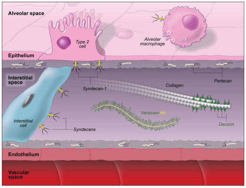
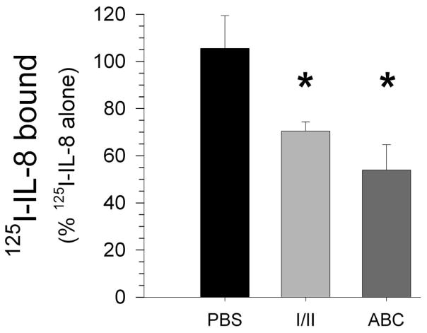
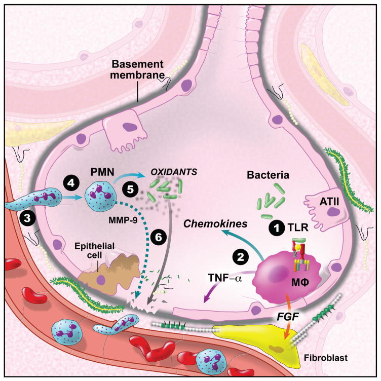
Exposure to viruses and bacteria results in lung infections and places a significant burden on public health. The innate immune system is an early warning system that recognizes viruses and bacteria, which results in the rapid production of inflammatory mediators such as cytokines and chemokines and the pulmonary recruitment of leukocytes. When leukocytes emigrate from the systemic circulation through the extracellular matrix (ECM) in response to lung infection they encounter proteoglycans, which consist of a core protein and their associated glycosaminoglycans. In this review, we discuss how proteoglycans serve to modify the pulmonary inflammatory response and leukocyte migration through a number of different mechanisms including: (1) The ability of soluble proteoglycans or fragments of glycosaminoglycans to activate Toll-like receptor (TLRs) signaling pathways; (2) The binding and sequestration of cytokines, chemokines, and growth factors by proteoglycans; (3) the ability of proteoglycans and hyaluronan to facilitate leukocyte adhesion and sequestration; and (4) The interactions between proteoglycans and matrix metalloproteinases (MMP) that alter the function of these proteases. In conclusion, proteoglycans fine-tune tissue inflammation through a number of different mechanisms. Clarification of the mechanisms whereby proteoglycans modulate the pulmonary inflammatory response will most likely lead to new therapeutic approaches to inflammatory lung disease and lung infection.


Figures

Figure 1
Figure 1
Proteoglycans found in normal lungs. Perlecan, a HSPG, is found in the basal lamina of epithelial and endothelial cells. The CSPGs, versican and decorin, are found in the interstitial space of the lungs. Versican binds to the glycosaminoglycan, hyaluronan, to form high-molecular weight complexes. Decorin binds to collagen and helps stabilize the collagen-elastin network. Syndecans are membrane proteoglycans that interact with matrix proteins. This figure is not meant to represent the concentrations of the different proteoglycans in normal lungs and is meant to only show their approximate location in the alveolus. (This figure is from a chapter by Frevert, et al., (Frevert and Wight, 2006) with permission)
Figure 2
Figure 2
The removal of the glycosaminoglycans heparan sulfate and chondroitin sulfate significantly decreases the low-affinity binding of 125I-CXCL8/IL-8 in lung tissue. Lung tissue was treated with heparinase I/II (I/II) and Chondroitinase ABC (ABC) prior to incubation of tissue with a trace amount of 125I-CXCL8/IL-8 [1 × 10⁻¹⁰ M] in combination with an excess of unlabeled CXCL8 [1 × 10⁻⁶ M]. The values are the mean + SEM (n = 4, *, p < 0.02). (This figure is from studies by Frevert, et al., (Frevert et al., 2003)with permission.)
Figure 3
Figure 3
In situ tissue-binding assay showing the binding of rhCXCL8/IL-8 to alveolar macrophages (arrows), endothelial cells (small arrowhead) and perivascular lymphatics (large arrowheads) around a pulmonary artery (A and B) and to the cell surface of airway epithelial cells (yellow arrow) and alveolar macrophage (white arrow) (C). rhCXCL8 is stained brown (A) in the bright field image and red in the confocal images (B and C). The negative control is a serial tissue section incubated with PBS instead of rhCXCL8 (D). Nuclei are stained with methylene green in the bright field images (A) and ToPro-3 (Blue) in the confocal images (B, C, D). Tissue autofluorescence (green) was used to improve the image quality of the confocal images (B – D). (This figure is from studies by Frevert, et al., (Frevert et al., 2003) with permission.)
Figure 4
Figure 4
An electron micrograph (EM) of normal lung tissue showing microdomains in the extracellular matrix which contain varying amounts of sulfation. Cytochemical visualization of sulfation in the extracellular matrix was performed using high iron diamine (HID), which binds to sulfates in lung tissue. The HID reaction product forms discrete 5–12 nm silver particles following appropriate intensification and is seen as discrete dark spots (Sannes, 1984). The basement membrane under a type I cell and an adjacent fibroblast (F) is highly sulfated (Arrows in 2A and 2B). The microdomains surrounding the pulmonary fibroblast show this cell to have microdomains of high and low sulfation. The extracellular matrix adjacent to the ATI cell is highly sulfated (white arrows in 2A and 2B) whereas the extracellular matrix adjacent to the endothelial cell (EC) is undersulfated (black arrowheads in 2A and 2B). In contrast to the ATI cell, this EM shows that the extracellular matrix underlying the type II cell (ATII) is undersulfated. White arrowheads in 2A and 2C define the extracellular matrix underlying the ATII cell and a pulmonary capillary EC (This figure is from studies by Frevert, et al., (Frevert and Sannes, 2006) with permission.).
Figure 5
Figure 5
Positive staining (brown) for syndecan-1 (syndecan-1) in normal mouse lungs shows a basal lateral distribution in airway epithelial cells and immunoreactivity in cells of the alveolar septa. Immunohistochemistry for syndecan-1 was performed with a rat anti-mouse syndecan-1 IgG (BD/Pharmingen, Franklin Lakes, NJ).
Figure 6
Figure 6
Syndecan-1/KC Complexes control the pulmonary recruitment of neutrophils in mice treated with bleomycin. (A) KC was immunoprecipitated (IP) from BAL of WT mice 1 day post-bleomycin. BAL and post-IP supernatants and pellets were electrophoresed and blotted for syndecan-1 ectodomain. The arrowhead indicates the band specifically coprecipitated with KC. (B) KC and syndecan-1 were immunoprecipitated from BAL of WT mice 1 day post-bleomycin. Two additional syndecan-1 IPs were done on the post-IP supernatant. The level of KC protein in the BAL and in the post-IP supernatants was quantified by ELISA. (C–E) WT and syndecan-1 null mice (SYN1−/−) (n = 3) were instilled with 0.15 U bleomycin. KC levels in BAL (C) and lung homogenates (D) were determined by ELISA. (E) Neutrophils (PMNs) in BAL were counted and expressed as a percent of total leukocytes. Data are the mean ± SE.. (This figure is from studies by Li, et al., (Li et al., 2002) with permission.)
Figure 7
Figure 7
The recognition of bacteria and viruses in the lungs results in the activation of Toll-like receptor (TLR) signalling pathways, which leads to pulmonary inflammation and under ideal conditions the clearance of the pathogen. Proteoglycans and/or their glycosaminoglycans modify the inflammatory response in lungs through a number of different mechanisms. (1) The release of soluble proteoglycans such as biglycan or degradation of glycosaminoglycan such as hyaluronan and heparan sulfate can activate TLRs. (2) Cytokines, chemokines, and growth factors bind to glycosaminoglycans, which can either increase or decrease their biological activity. (3) Adhesion molecules including the selectins, integrins, and CD44 bind to proteoglycans and glycosaminoglycan, which suggests that these proteins play a critical role in leukocyte adhesion and migration. (4) Chemokine-glycosaminoglycan interactions provide fine-tune control of chemokine-gradient formation and leukocyte migration in tissue. (5) Activation of stromal and immune cells results in the release of MMP. Growing evidence shows that interactions between proteoglycans and MMPs play important roles in the regulation of the innate immune response. (6) Degradation of proteoglycans by MMPs and other proteases controls the amount and localization of proteoglycans in lungs. In addition, proteolytic cleavage of proteoglycans leads to the unmasking of cryptic fragments.

References

    1. Adair-Kirk TL, Senior RM. Fragments of extracellular matrix as mediators of inflammation. Int J Biochem Cell Biol. 2008;40:1101–1110. - PMC - PubMed
    1. Akira S. Innate immunity to pathogens: diversity in receptors for microbial recognition. Immunol Rev. 2009;227:5–8. - PubMed
    1. Ali S, Palmer AC, Banerjee B, Fritchley SJ, Kirby JA. Examination of the Function of RANTES, MIP-1alpha, and MIP-1beta following Interaction with Heparin-like Glycosaminoglycans. J Biol Chem. 2000;275:11721–11727. - PubMed
    1. Allen BL, Filla MS, Rapraeger AC. Role of heparan sulfate as a tissue-specific regulator of FGF-4 and FGF receptor recognition. J Cell Biol. 2001;155:845–858. - PMC - PubMed
    1. Amara A, Lorthioir O, Valenzuela A, Magerus A, Thelen M, Montes M, Virelizier JL, Delepierre M, Baleux F, Lortat-Jacob H, Arenzana-Seisdedos F. Stromal cell-derived factor-1alpha associates with heparan sulfates through the first beta-strand of the chemokine. J Biol Chem. 1999;274:23916–23925. - PubMed

LinkOut - more resources