. 2010 May 28;16(20):2504-19.
doi: 10.3748/wjg.v16.i20.2504.

Clinicopathological features and prognosis assessment of extranodal follicular dendritic cell sarcoma

Affiliations

Clinicopathological features and prognosis assessment of extranodal follicular dendritic cell sarcoma

Lan Li et al. World J Gastroenterol. .

Abstract

Aim: To establish a model for prognosis assessment of extranodal follicular dendritic cell (FDC) sarcoma.

Methods: Nine lesions were examined by routine and molecular approaches. Clinicopathological factors from the new cases and 97 reported cases were analyzed for their prognostic values.

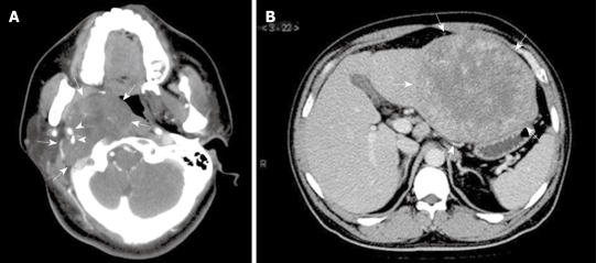
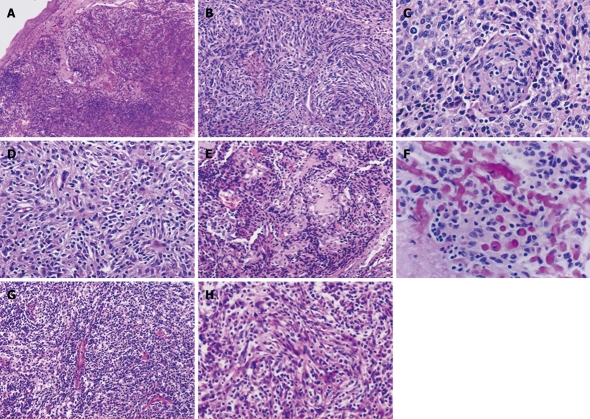
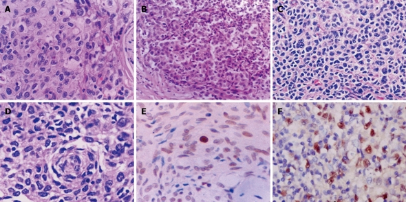
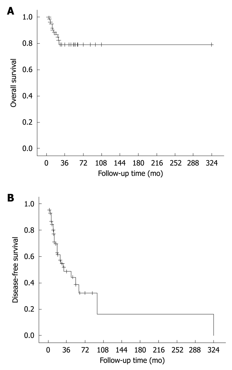
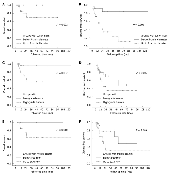
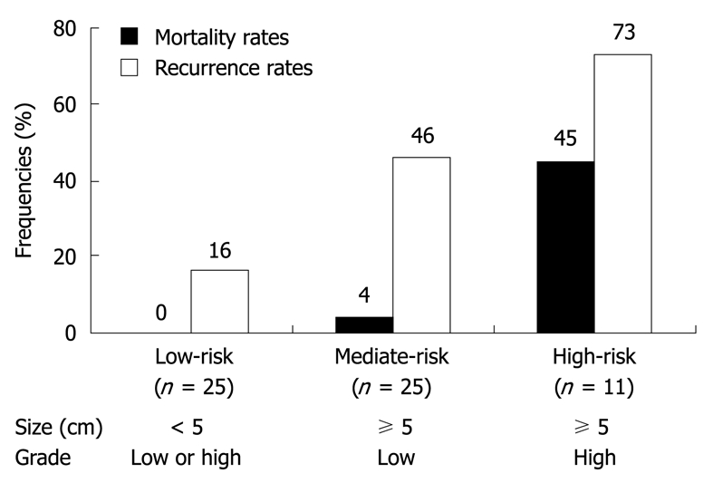
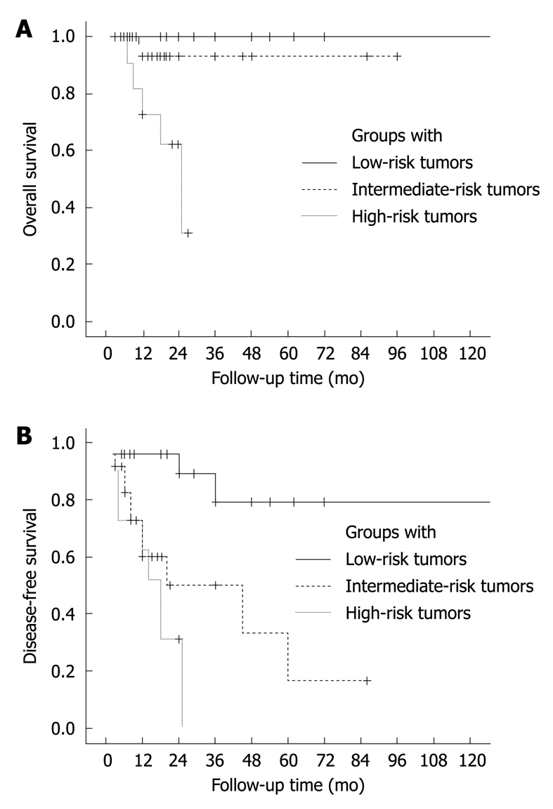
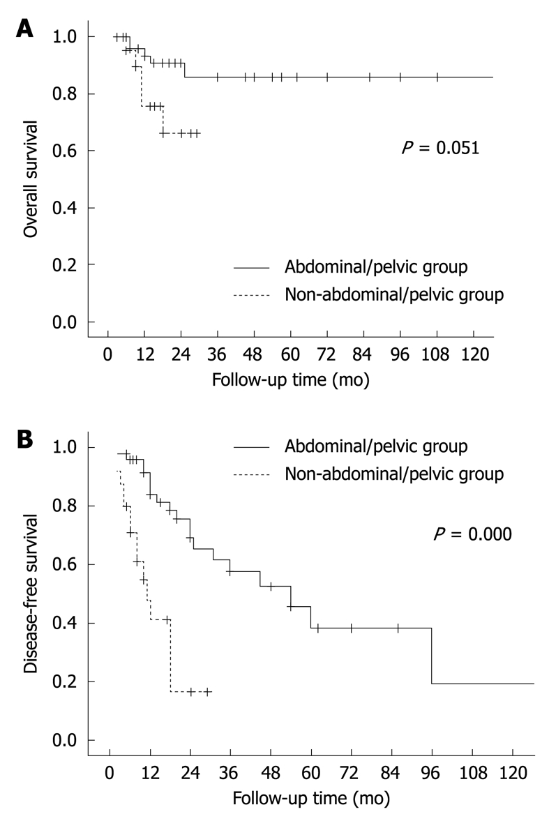
Results: The current lesions were found in five male and four female patients, located mainly in the head and neck area and averaging 7.2 cm in size. Six patients had recurrence or metastasis and three remained free of disease. The 106 patients (male/female ratio, 1.1:1) were aged from 9 to 82 years (median, 44 years). The tumor sizes ranged from 1.5 to 21 cm (mean, 7.4 cm). Abdominal/pelvic region was affected most frequently (43%). Surgical resection was performed in 100 patients, followed by radiation and/or chemotherapy in 35 of them. Follow-up data were available in 91 cases, covering a period of 3-324 mo (mean, 27 mo; median, 19 mo). Of the informative cases, 38 (42%) had recurrence or metastasis, and 12 (13%) died of the disease. These tumors were classified histologically into low- and high-grade lesions. A size > or = 5 cm (P = 0.003), high-grade histology (P = 0.046) and a mitotic count > or = 5/10 HPF (P = 0.013) were associated with tumor recurrence. The lesions were defined as low-, intermediate- and high-risk tumors, and their recurrence rates were 16%, 46% and 73%, and their mortality rates 0%, 4% and 45%, respectively.

Conclusion: Extranodal FDC tumors behave like soft tissue sarcomas. Their clinical outcomes are variable and can be evaluated according to their sizes and grades.


Figures

Figure 1
Figure 1
Radiological features of extranodal follicular dendritic cell (FDC) sarcoma shown by computed tomographic scan. A: Case 8, a tumor (arrows) at the right parapharyngeal space showing soft tissue-like density and an expansive growth pattern, with the internal and external carotid arteries (arrowheads) engulfed; B: Case 6, a well-circumscribed mass (arrows) at the left lobe of liver, showing irregular enhancement at its periphery.
Figure 2
Figure 2
Typical features of follicular dendritic cell sarcoma of the conventional (A-F) and inflammatory pseudotumor-like types (G and H). Spindle and ovoid tumor cells, frequently growing in nodules as in Case 1 (A), arrange in whorl (Case 3, B and C) and storiform (Case 3, D) and fascicular patterns (Case 6, H), with sprinkling small lymphocytes throughout the former type of lesions and numerous plasma cells and lymphocytes in the latter. Perivascular sclerosis was noted in Case 3 (E), with foci of osteoid matrix deposition resembling osteosarcoma (F). HE: A, × 40; B, E and G, × 100; C, D, F and H, × 200.
Figure 3
Figure 3
Atypical morphology of FDC sarcoma (A-D) and expression of p53 protein (E) and Epstein-Barr virus-encoded RNA (EBER) (F) in tumor cells. A-D: Epithelioid (A and B) and pleomorphic tumor cells (C and D) are arranged in a sheet-like or diffuse pattern. Lymphocyte infiltration is less prominent (A and B) or absent (C and D) in these areas. HE: A, C and D, × 400; B, × 200; E: Nuclear immunoreactivity for p53 protein in majority of tumor cells. S-P, × 400; F: In situ hybridization signal for EBER in tumor cells, × 400.
Figure 4
Figure 4
Overall (A) and disease-free survival curves (B) of FDC sarcoma. The results are based on follow-up data of 91 informative cases.
Figure 5
Figure 5
Overall (A, C and E) and disease-free survival curves (B, D and F) of patients with FDC sarcoma, showing data of 120 mo.
Figure 6
Figure 6
Definitions of low- (low-risk), intermediate- (mediate-risk) and high-risk groups (high-risk) of extranodal FDC sarcomas. Mortality rates: overall, P = 0.000; low-risk vs mediate-risk, P = 1.000; low-risk vs high-risk, P = 0.001; mediate-risk vs high-risk, P = 0.006; Recurrence rates: overall, P = 0.001; low-risk vs mediate-risk, P = 0.032; low-risk vs high-risk, P = 0.002; mediate-risk vs high-risk, P = 0.167.
Figure 7
Figure 7
Overall (A) and disease-free survival curves (B) of patients with FDC sarcoma, estimated in groups with low-, intermediate- and high-risk tumors, showing data of 120 mo. A: Overall, P = 0.000; low-risk vs mediate-risk, P = 0.273; low-risk vs high-risk, P = 0.000; mediate-risk vs high-risk, P = 0.017; B: Overall, P = 0.000; low-risk vs mediate-risk, P = 0.003; low-risk vs high-risk, P = 0.000; mediate-risk vs high-risk, P = 0.207.
Figure 8
Figure 8
Overall (A) and disease-free survival curves (B) of abdominal/pelvic FDC sarcomas. The lesions are divided into hepatic and extrahepatic groups.
Figure 9
Figure 9
Overall (A) and disease-free survival curves (B) of patients with extrahepatic FDC sarcomas, showing data of 120 mo. The lesions are divided into abdominal/pelvic and non-abdominal/pelvic groups.

References

    1. Monda L, Warnke R, Rosai J. A primary lymph node malignancy with features suggestive of dendritic reticulum cell differentiation. A report of 4 cases. Am J Pathol. 1986;122:562–572. - PMC - PubMed
    1. Pileri SA, Grogan TM, Harris NL, Banks P, Campo E, Chan JK, Favera RD, Delsol G, De Wolf-Peeters C, Falini B, et al. Tumours of histiocytes and accessory dendritic cells: an immunohistochemical approach to classification from the International Lymphoma Study Group based on 61 cases. Histopathology. 2002;41:1–29. - PubMed
    1. Chan JK, Fletcher CD, Nayler SJ, Cooper K. Follicular dendritic cell sarcoma. Clinicopathologic analysis of 17 cases suggesting a malignant potential higher than currently recognized. Cancer. 1997;79:294–313. - PubMed
    1. Cheuk W, Chan JK, Shek TW, Chang JH, Tsou MH, Yuen NW, Ng WF, Chan AC, Prat J. Inflammatory pseudotumor-like follicular dendritic cell tumor: a distinctive low-grade malignant intra-abdominal neoplasm with consistent Epstein-Barr virus association. Am J Surg Pathol. 2001;25:721–731. - PubMed
    1. Chen TC, Kuo TT, Ng KF. Follicular dendritic cell tumor of the liver: a clinicopathologic and Epstein-Barr virus study of two cases. Mod Pathol. 2001;14:354–360. - PubMed

Publication types

MeSH terms

Substances