

A case of intrahepatic clear cell cholangiocarcinoma
- PMID: 20503460
- PMCID: PMC2877190
- DOI: 10.3748/wjg.v16.i20.2571
A case of intrahepatic clear cell cholangiocarcinoma
Abstract
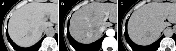
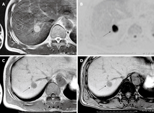
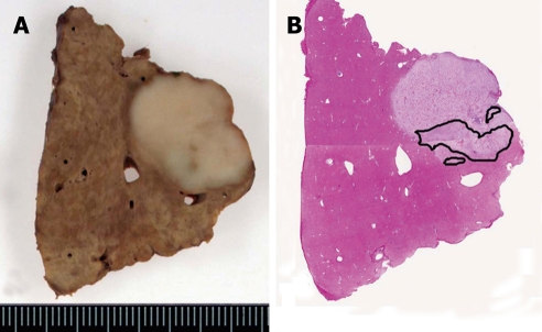
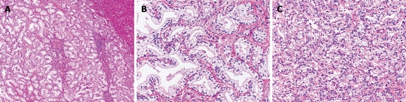
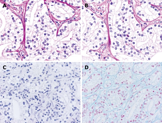
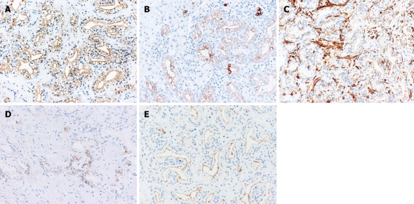
Intrahepatic clear cell cholangiocarcinoma is very rare - only 8 cases have been reported. A 56-year-old Japanese man with chronic hepatitis B infection was diagnosed with a 2.2 cm hepatocellular carcinoma on imaging, and hepatic segmentectomy was performed. Histopathologically, the tumor cells had copious clear cytoplasm and formed glandular structures or solid nests. These pathological findings suggested the tumor was a clear cell variant of intrahepatic cholangiocarcinoma. Particular stains and radiological images suggested that the cause of the clear cell change had been glycogen, not mucin nor lipid. On immunohistochemical staining, cytokeratin (CK) 7 and CK19 were positive, whereas CK20 was negative. Vimentin was detected on the cell membranes, and CD56 was focally positive. The patient was given adjuvant chemotherapy and is currently free from the tumor 7 mo postoperatively. Careful follow-up with adequate postoperative supplementary chemotherapy is necessary because the characteristics of this type of tumor are unknown.
Figures
Similar articles
-
Intrahepatic clear cell cholangiocarcinoma: immunohistochemical aspects in a very rare type of cholangiocarcinoma.Am J Surg Pathol. 2007 Jun;31(6):902-6. doi: 10.1097/PAS.0b013e31802c0c8a. Am J Surg Pathol. 2007. PMID: 17527078
-
Intrahepatic clear cell cholangiocarcinoma - An uncommon histologic subtype: case report and literature review.Rev Esp Enferm Dig. 2017 May;109(5):382-385. doi: 10.17235/reed.2017.4239/2016. Rev Esp Enferm Dig. 2017. PMID: 28155327 Review.
-
[Expression of mucin glycoproteins and cytokeratins in intrahepatic cholangiocarcinoma].Zhonghua Bing Li Xue Za Zhi. 2008 Nov;37(11):749-53. Zhonghua Bing Li Xue Za Zhi. 2008. PMID: 19094709 Chinese.
-
Immunohistochemical staining in the diagnosis of pancreatobiliary and ampulla of Vater adenocarcinoma: application of CDX2, CK17, MUC1, and MUC2.Am J Surg Pathol. 2005 Mar;29(3):359-67. doi: 10.1097/01.pas.0000149708.12335.6a. Am J Surg Pathol. 2005. PMID: 15725805
-
Synchronous double cancers of primary hepatocellular carcinoma and intrahepatic cholangiocarcinoma: a case report and review of the literature.World J Surg Oncol. 2014 Nov 10;12:337. doi: 10.1186/1477-7819-12-337. World J Surg Oncol. 2014. PMID: 25385169 Free PMC article. Review.
Cited by
-
Intrahepatic cholangiocarcinoma with clear cell type following laparoscopic curative surgery.Surg Case Rep. 2020 Oct 7;6(1):264. doi: 10.1186/s40792-020-01041-2. Surg Case Rep. 2020. PMID: 33026548 Free PMC article.
-
Cytopathologic, histopathologic, and immunohistochemical features of intrahepatic clear cell bile duct adenoma: a case report and review of the literature.Case Rep Pathol. 2014;2014:874826. doi: 10.1155/2014/874826. Epub 2014 May 19. Case Rep Pathol. 2014. PMID: 24955270 Free PMC article.
-
A case of primary clear cell hepatocellular carcinoma in a non-cirrhotic liver: an immunohistochemical and ultrastructural study.Rare Tumors. 2012 Apr 12;4(2):e29. doi: 10.4081/rt.2012.e29. Epub 2012 May 17. Rare Tumors. 2012. PMID: 22826786 Free PMC article.
-
Clear Cell Carcinoma: A Rare Variant of Cholangiocarcinoma Case Report and Systematic Review.Case Rep Gastrointest Med. 2025 Jun 28;2025:1716741. doi: 10.1155/crgm/1716741. eCollection 2025. Case Rep Gastrointest Med. 2025. PMID: 40620746 Free PMC article.
-
Rare Histological Variants of Liver Cancer and Their Management: A Single-Institution Experience.Case Reports Hepatol. 2021 Apr 21;2021:6654229. doi: 10.1155/2021/6654229. eCollection 2021. Case Reports Hepatol. 2021. PMID: 33968454 Free PMC article.
References
-
- Nakanuma Y, Sripa B, Vatanasapt V, Leong AS-Y, Ponchon T, Ishak KG. Intrahepatic cholangiocarcinoma. In: Hamilton SR, Aaltonen LA, editors. World Health Organization Classification of Tumours, Pathology&Genetics: Tumours of the Digestive System. Lyon: IARC Press; 2000. pp. 173–180.
-
- Kitajima K, Shiba H, Nojiri T, Uwagawa T, Ishida Y, Ichiba N, Yanaga K. Intrahepatic cholangiocarcinoma mimicking hepatic inflammatory pseudotumor. J Gastrointest Surg. 2007;11:398–402. - PubMed
-
- Haas S, Gütgemann I, Wolff M, Fischer HP. Intrahepatic clear cell cholangiocarcinoma: immunohistochemical aspects in a very rare type of cholangiocarcinoma. Am J Surg Pathol. 2007;31:902–906. - PubMed
-
- Ishak KG, Goodman ZD, Stocker JT. Atlas of tumor pathology third series fascicle 31: tumors of the liver and intrahepatic bile ducts. Washington, DC: Armed Forces Institute of Pathology; 1999. pp. 245–263.
Publication types
MeSH terms
Substances
LinkOut - more resources
Full Text Sources
Research Materials
