. 2010 Mar 15;1(1):5.
doi: 10.1186/scrt5.

Immunomodulatory properties of stem cells from human exfoliated deciduous teeth

Affiliations

Immunomodulatory properties of stem cells from human exfoliated deciduous teeth

Takayoshi Yamaza et al. Stem Cell Res Ther. .

Abstract

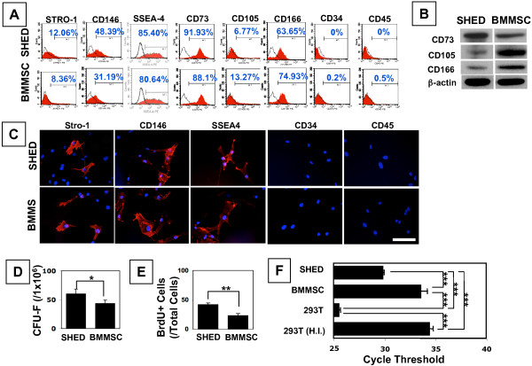
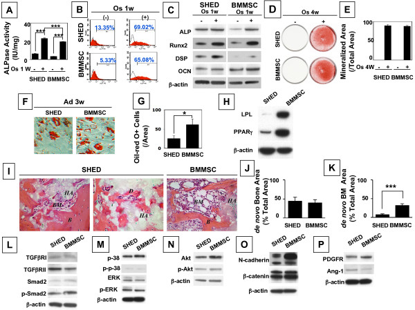
Introduction: Stem cells from human exfoliated deciduous teeth (SHED) have been identified as a population of postnatal stem cells capable of differentiating into osteogenic and odontogenic cells, adipogenic cells, and neural cells. Herein we have characterized mesenchymal stem cell properties of SHED in comparison to human bone marrow mesenchymal stem cells (BMMSCs).

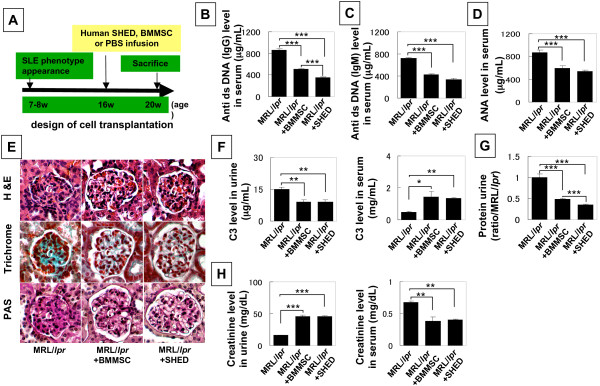
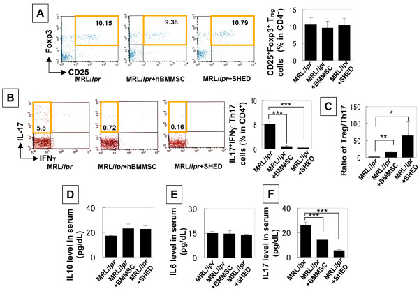
Methods: We used in vitro stem cell analysis approaches, including flow cytometry, inductive differentiation, telomerase activity, and Western blot analysis to assess multipotent differentiation of SHED and in vivo implantation to assess tissue regeneration of SHED. In addition, we utilized systemic SHED transplantation to treat systemic lupus erythematosus (SLE)-like MRL/lpr mice.

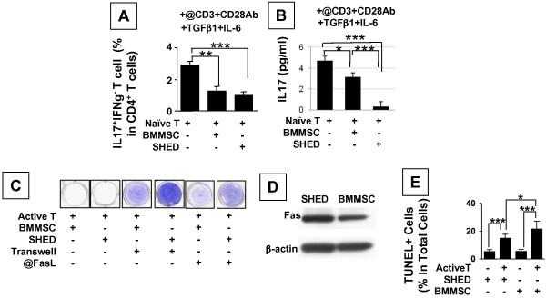
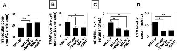
Results: We found that SHED are capable of differentiating into osteogenic and adipogenic cells, expressing mesenchymal surface molecules (STRO-1, CD146, SSEA4, CD73, CD105, and CD166), and activating multiple signaling pathways, including TGFbeta, ERK, Akt, Wnt, and PDGF. Recently, BMMSCs were shown to possess an immunomodulatory function that leads to successful therapies for immune diseases. We examined the immunomodulatory properties of SHED in comparison to BMMSCs and found that SHED had significant effects on inhibiting T helper 17 (Th17) cells in vitro. Moreover, we found that SHED transplantation is capable of effectively reversing SLE-associated disorders in MRL/lpr mice. At the cellular level, SHED transplantation elevated the ratio of regulatory T cells (Tregs) via Th17 cells.

Conclusions: These data suggest that SHED are an accessible and feasible mesenchymal stem cell source for treating immune disorders like SLE.


Figures

Figure 1
Figure 1
Characterization of SHED in comparison to BMMSCs. (A) Flow cytometric analysis of cultured SHED at passage 3 revealed expression of STRO-1 (12.06%), CD146 (48.39%), SSEA4 (85.40%), CD73 (91.93%), CD105 (6.77%), CD166 (63.65%), but was negative for surface molecules CD34 and CD45. SHED express high levels of STRO-1 and CD146 (n = 5; P < 0.05) and low level of CD105 (n = 5; P < 0.01) compared to expression levels of STRO-1 (8.36%), CD146 (31.19%), and CD105 (13.27%) in BMMSCs. These signals are shown as the red area. Solid lines indicate signals for isotype matched control antibodies. M1 window show the positive expression defined as the level of fluorescence greater than 99% of the corresponding isoytpe-matched control antibodies. Representative histograms are shown among five donors. (B) Immunoblot analysis confirmed expression of CD73, CD105 and CD166 in SHED and BMMSCs. Representative images of n = 5 donors are presented as results. (C) Immunofluoresence confirmed that SHED express STRO-1, CD146, and SSEA4 along with negative for CD34 and CD45. Red fluorescence indicates the expression of cell surface markers. Blue cell nuclei were stained by DAPI. Images were representative data of independent experiment (n = 5) with consistent results (Bar = 50 μm). (D) SHED were able to form significantly high numbers of single colonies than BMMSCs when 1 × 10⁶ cells were plated at a low density (*P < 0.05) and cultured for 10 days. (E) The proliferation rates of SHED and BMMSCs were assessed by co-culture with BrdU for 18 hours. The number of BrdU-positive cells was presented as a percentage of the total number of cells counted from five replicate cultures. SHED showed a significantly higher proliferation rate in comparison to BMMSCs (**P < 0.01). (F) SHED showed a high activity of telomerase compared to BMMSCs assessed by real time PCR. HEK293T cells (239T) were used as a positive control and heat inactivated 293T (H.I.) cells were used as a negative control. The activity was indicated by a PCR cycle threshold and averaged from three replicated cultures (***P < 0.001).
Figure 2
Figure 2
Mesenchymal stem cell properties of SHED. (A-E) SHED showed a similar osteogenic differentiation potential to BMMSCs. After one week culture induction under osteogenic conditions, ALP activity and numbers of ALP positive cells in SHED and BMMSCs were significantly higher than that of the control SHED and BMMSCs, respectively, by ALP staining (Representative of n = 5) (A) and flow cytometric analysis (Representative of n = 3) (B). Meanwhile, immunoblot analysis showed that the osteogenic induction elevates expression levels of ALP, Runx2, DSP, and OCN in SHED and BMMSCs (C) (***P < 0.001, n = 5). β-actin was used as an internal control. After four weeks culture induction in osteogenic medium, SHED showed increased capacity of forming mineralized nodules as assessed by alizarin red staining (Representative of n = 5). (D). Alizarin red-positive area corresponding to total area was averaged from five independent groups (E). (F-H) SHED showed reduced potential of differentiating into adipocytes compared to BMMSCs. Three weeks post adipogenic induction, lipid accumulation in SHED was less than that in BMMSCs by Oil-red O staining (Representative of n = 5). (F). Number of oil-red O-positive (Oil-Red-O+) cells was calculated as a percentage to total cells and averaged from five independent cultures (G) (*P < 0.05). Immunoblot assay indicated that SHED expressed lower levels of adipocyte-specific molecules LPL and PPARγ than BMMSCs at three weeks post adipogenic culture (H). Three independent assays showed the similar results. (I-K) SHED were capable of forming mineralized tissue when transplanted subcutaneously into immunocompromised mice using HA/TCP as a carrier (Representative of n = 3). (I). It appeared that SHED form similar amounts of mineralized tissue as seen in a BMMSC transplant (Representative of n = 3) (I, J), but they generated significantly less bone marrow elements than BMMSCs (K). Newly formed mineralized tissue and bone marrow areas were calculated as a percentage of the total area and averaged from three independent transplant assays (***P < 0.001). B = bone, BM = bone marrow, C =: connective tissue, H =: hydroxyapatite and tricalcium carrier. (L-P) SHED and BMMSCs express multiple signaling pathways during culture expansion at passage 3. SHED and BMMSCs expressed TGFβ receptor I and II, Smad 2 and phosphorylated Smad 2 (L); P38, phosphorylated P38, ERK, and phosphorylated ERK (M); Akt and phosphorylated Akt (N); N-cadherin and β-catenin (O); PDGF receptor and Ang-1 (P). Representative image of n = 5.
Figure 3
Figure 3
SHED interplay with T-lymphocytes. (A, B) Under the anti-CD3 and CD28 antibody along with TGFβ1 and IL-2 stimulation, SHED showed a significant effect in reducing Th17 cell levels as seen in BMMSCs (A), however, SHED exhibited a significant capacity of inhibiting IL17 levels than BMMSCs (B) (n = 3, *P < 0.05,***P < 0.001). (C) PBMNCs activated by anti-CD3 antibody (@CD3Ab, 1 μg/ml) were capable of inducing significant SHED and BMMSC death as shown by toulidin blue staining. When cells were cultured in an indirect co-culture system using Transwell, activated slpenocytes they failed to induce SHED and BMMSC death. Neutralization with anti-FasL antibody (@FasLAb, 1 μg/ml) blocked PBMNC-induced SHED and BMMSC death. Representative of n = 3. (D) SHED express a higher level of Fas in comparison to that in BMMSCs by immunoblotting. Three independent experiments showed similar results. Representative of n = 3. (E) SHED death caused by active PBMNCs is through an apoptotic pathway according to the TUNEL staining. The SHED death rate was similar to BMMSCs. The percentage of TUNEL-positive (TUNEL+) nuclei was indicated to the total number of MSCs and averaged from five replicated cultures (***P < 0.005).
Figure 4
Figure 4
SHED transplantation reduced levels of autoantibodies and improved renal function in MRL/lpr mice. Figure 4A shows the scheme of SHED and BMMSC transplantation procedures. (B-D) ELISA quantified that levels of anti dsDNA IgG (B), IgM (C) and nuclear (D) antibodies (ANA) (mean ± SD) were significantly reduced in the peripheral blood of SHED and BMMSC treated MRL/lpr mice (n = 6) when compared to un-treated MRL/lpr mice c (n = 6) (***P < 0.001). It appeared that SHED transplantation resulted in a more significant reduction in anti IgG when compared to BMMSC transplantation (B). (E) MRL/lpr mice showed renal disorders such as nephritis with glomerular basal membrane disorder and mesangium cell over-growth. SHED and BMSSC transplantation resulted in a reduced basal membrane disorder and mesangium cell over-growth in glomerular (G) (upper panels, H&E staining; middle panels, trichrome staining; lower panels, periodic acid-schiff staining). Representative images of un-treated, SHED and BMMSC MRL/lpr (n = 6). (F) ELISA analysis showed that SHED transplantation has the same effect as seen in BMMSC transplantation in significantly reducing C3 level in urine and elevating C3 level in serum (n = 6, *P < 0.05, **P < 0.01). (G) SHED transplantation significantly reduced urine protein levels (mean ± SD) compared to BMMSC transplanted MRL/lpr mice (n = 6). ([[[P < 0.001). (H) Markedly increased urine creatinine and reduced serum creatinine were observed in SHED and BMMSC transplanted MRL/lpr mice (n = 6) compared to un-treated MRL/lpr mice (n = 6, [[[P < 0.001, [[P < 0.01).
Figure 5
Figure 5
The ratio of Tregs and Th17 cells may contribute to SHED mediated treatment in MRL/lpr mice. (A-C) Flow cytometric analysis showed that the number of CD25+Foxp3+ Tregs in CD4+ T lymphocytes of MRL/lpr spleen was not significantly changed in SHED and BMMSC transplantation (A). In contrast, SHED and BMMSC transplantation were capable of significantly reduced levels of CD4+IL17+ cells in spleen as compared to un-treated MRL/lpr mice (B). SHED transplantation significantly increased the ratio of Tregs and Th17 cells when compared to BMMSC transplantation group (C) ([[[P < 0.001, [[P < 0.01, [P < 0.05). Results were shown as mean ± SD from un-treated, SHED and BMMSC MRL/lpr (n = 6). (D-F) Although SHED and BMMSC transplantations failed to alter IL10 (D) and IL6 (E) levels in serum of MRL/lpr mice, IL17 levels were significantly down-regulated in SHED and BMMSC transplanted group compared to un-treated MRL/lpr mice (F). Results were shown as means ± SD from un-treated, SHED and BMMSC MRL/lpr (n = 6).
Figure 6
Figure 6
SHED transplantation reconstructed trabecular bone and inhibited osteoclast activity. (A) SHED transplantation showed the same effect in regenerating trabecular bone as seen in BMMSC transplanted MRL/lpr mice (n = 6) ([[P < 0.01). (B) TRAP staining showed that the number of TRAP positive osteoclasts was significantly reduced in SHED and BMMSC transplanted mice (n = 6, [P < 0.05). (C, D) ELISA revealed that SHED and BMMSC transplantations were capable of significantly reducing the levels (mean ± SD) of soluble RANKL (sRANKL) (C) and C-terminal telopeptides of type I collagen (CTX) (D) in serum of MRL/lpr mice (n = 6)(*P < 0.05, **P < 0.01).

Similar articles

Cited by

References

    1. Prockop DJ. Marrow stromal cells as stem cells for nonhematopoietic tissues. Science. 1997;276:71–74. doi: 10.1126/science.276.5309.71. - DOI - PubMed
    1. Bianco P, Riminucci M, Gronthos S, Robey PG. Bone marrow stromal stem cells: nature, biology, and potential applications. Stem Cells. 2001;19:180–192. doi: 10.1634/stemcells.19-3-180. - DOI - PubMed
    1. Friedenstein AJ, Chailakhyan RK, Latsinik NV, Panasyuk AF, Keiliss-Borok IV. Stromal cells responsible for transferring the microenvironment of the hemopoietic tissues. Cloning in vitro and retransplantation in vivo. Transplantation. 1974;17:331–340. doi: 10.1097/00007890-197404000-00001. - DOI - PubMed
    1. Owen M, Friedenstein AJ. Stromal stem cells: marrow-derived osteogenic precursors. Ciba Found Symp. 1988;136:42–60. - PubMed
    1. Pittenger MF, Mackay AM, Beck SC, Jaiswal RK, Douglas R, Mosca JD, Moorman MA, Simonetti DW, Craig S, Marshak DR. Multilineage potential of adult human mesenchymal stem cells. Science. 1999;284:143–147. doi: 10.1126/science.284.5411.143. - DOI - PubMed

Publication types

MeSH terms