

Subcellular localization of Mayven following expression of wild type and mutant EGFP tagged cDNAs
- PMID: 20504342
- PMCID: PMC2901378
- DOI: 10.1186/1471-2202-11-63
Subcellular localization of Mayven following expression of wild type and mutant EGFP tagged cDNAs
Abstract
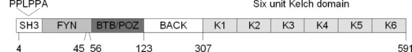
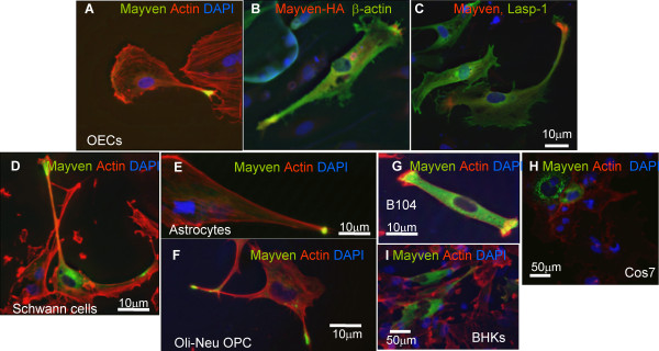
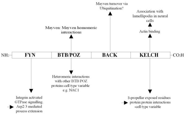
Background: Process formation by glial cells is crucial to their function. Mayven, an actin binding, multi-domain polypeptide, and member of the BTB-BACK-Kelch family have been shown to be important in oligodendrocyte process extension. To assess the role of Mayven in neural cell process extension we have tracked the subcellular distribution of exogenous Mayven following expression of a rat Mayven -EGFP cDNA in a variety of neural cell backgrounds and specifically in OEC tranfectants following drug treatment to disrupt the integrity of the cytoskeleton. A comparison was made between the subcellular localization following transient transfection of OECs with full-length Mayven cDNA and a series of mutant domain constructs.
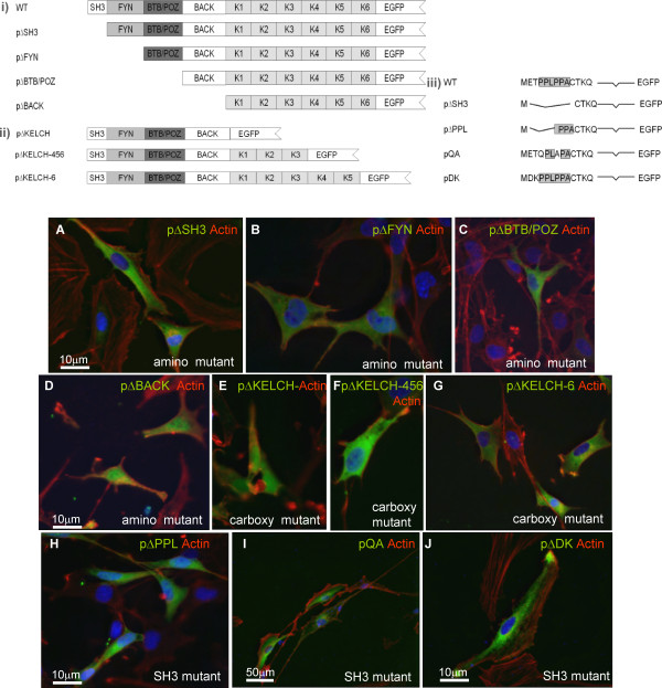
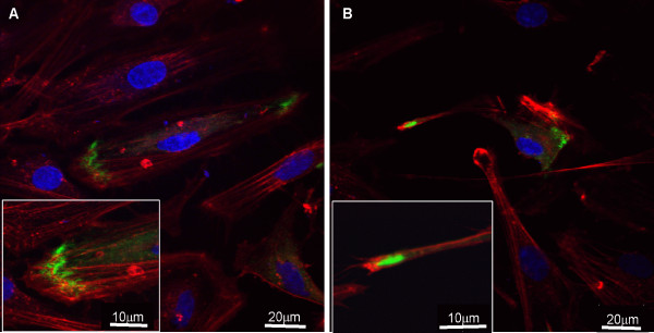
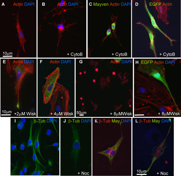
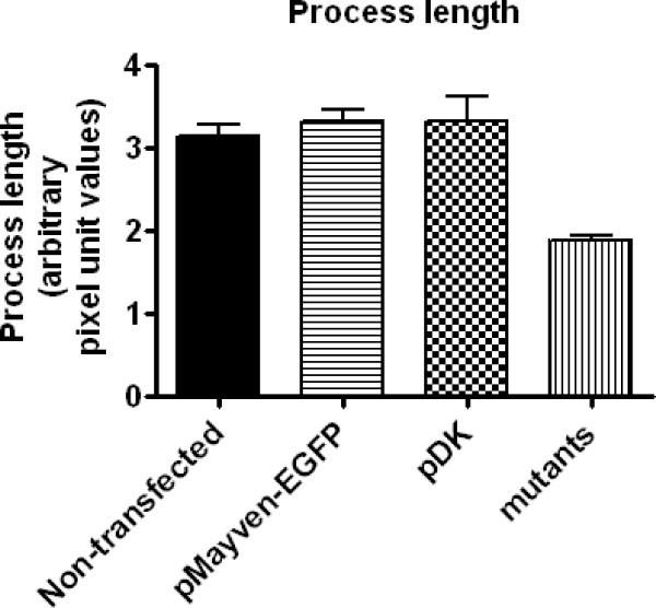
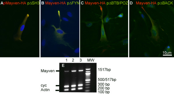
Results: The subcellular location of Mayven in OEC transfectants showed a characteristic distribution with intense foci of staining towards the process tips corresponding to regions of accumulated Mayven overlapping in part with lammelipodial actin and was absent from the filipodia and the outer membrane. This signature pattern was also observed in Schwann cells, Oli-Neu cells, astrocytes and the neuroblastoma cell line B104 transfectants and resembled the exogenous and endogenous Mayven distribution in oligodendrocytes. This contrasted with the localization pattern in non-neural cells. There was a re-localization of Mayven in OEC transfectants following drug treatment to challenge the integrity of the actin cytoskeleton while breakdown of the microtubular component had no discernible impact on the accumulation of Mayven in the process tips. Deletion of the first three amino acids of the SH3 motif of the putative Fyn Kinase binding domain at the amino terminus significantly compromised this signature pattern as did the removal of the last Kelch repeat unit of six unit Kelch domain comprising the carboxyl terminus. In addition, there was a reduction in process length in mutant transfectants. Co-expression studies with a haemagglutinin (HA) tagged wild type Mayven cDNA and EGFP tagged mutant cDNAs suggested a homomeric interaction mediated by the BTB/POZ domain.
Conclusions: Exogenous Mayven is transported to the lamellipodia in neural transfectants associating with the actin cytoskeletal network. In addition to the importance of the internal BTB/POZ domain, this subcellular distribution pattern is dependent on the presence of an intact amino and carboxyl terminus.
Figures
Similar articles
-
Characterization of Mayven, a novel actin-binding protein predominantly expressed in brain.Mol Biol Cell. 1999 Jul;10(7):2361-75. doi: 10.1091/mbc.10.7.2361. Mol Biol Cell. 1999. PMID: 10397770 Free PMC article.
-
Role of Mayven, a kelch-related protein in oligodendrocyte process formation.J Neurosci Res. 2005 Sep 1;81(5):622-31. doi: 10.1002/jnr.20588. J Neurosci Res. 2005. PMID: 16035103
-
Process elongation of oligodendrocytes is promoted by the Kelch-related actin-binding protein Mayven.J Neurochem. 2005 Mar;92(5):1191-203. doi: 10.1111/j.1471-4159.2004.02946.x. J Neurochem. 2005. PMID: 15715669
-
Mayven induces c-Jun expression and cyclin D1 activation in breast cancer cells.Oncogene. 2005 Mar 31;24(14):2398-409. doi: 10.1038/sj.onc.1208466. Oncogene. 2005. PMID: 15735724
-
The BACK domain in BTB-kelch proteins.Trends Biochem Sci. 2004 Dec;29(12):634-7. doi: 10.1016/j.tibs.2004.10.003. Trends Biochem Sci. 2004. PMID: 15544948 Review.
Cited by
-
In vivo electroporation and non-protein based screening assays to identify antibodies against native protein conformations.Hybridoma (Larchmt). 2011 Oct;30(5):409-18. doi: 10.1089/hyb.2010.0120. Hybridoma (Larchmt). 2011. PMID: 22008067 Free PMC article.
-
Update on the Kelch-like (KLHL) gene family.Hum Genomics. 2013 May 15;7(1):13. doi: 10.1186/1479-7364-7-13. Hum Genomics. 2013. PMID: 23676014 Free PMC article.
-
Interaction of an intracellular pentraxin with a BTB-Kelch protein is associated with ubiquitylation, aggregation and neuronal apoptosis.Mol Cell Neurosci. 2011 Aug;47(4):254-64. doi: 10.1016/j.mcn.2011.04.005. Epub 2011 Apr 28. Mol Cell Neurosci. 2011. PMID: 21549840 Free PMC article.
-
The innate immunity adaptor SARM translocates to the nucleus to stabilize lamins and prevent DNA fragmentation in response to pro-apoptotic signaling.PLoS One. 2013 Jul 29;8(7):e70994. doi: 10.1371/journal.pone.0070994. Print 2013. PLoS One. 2013. PMID: 23923041 Free PMC article.
References
Publication types
MeSH terms
Substances
LinkOut - more resources
Full Text Sources
Molecular Biology Databases
Research Materials
Miscellaneous
