

Anti-proliferative effect of LXR agonist T0901317 in ovarian carcinoma cells
- PMID: 20504359
- PMCID: PMC2890636
- DOI: 10.1186/1757-2215-3-13
Anti-proliferative effect of LXR agonist T0901317 in ovarian carcinoma cells
Abstract
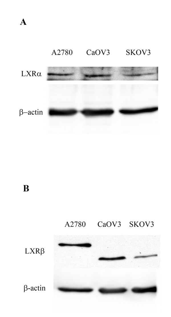
Background: Ovarian cancer is the most common cause of cancer related death from gynecologic tumors in the United States. The insidious nature of the disease precludes early diagnosis, therefore surgical debulking and chemotherapy are considered as standard treatment modalities for advanced stages. We investigated the effect of the LXR agonist, T0901317, on ovarian cancer cell proliferation and apoptosis as a potential therapeutic agent.
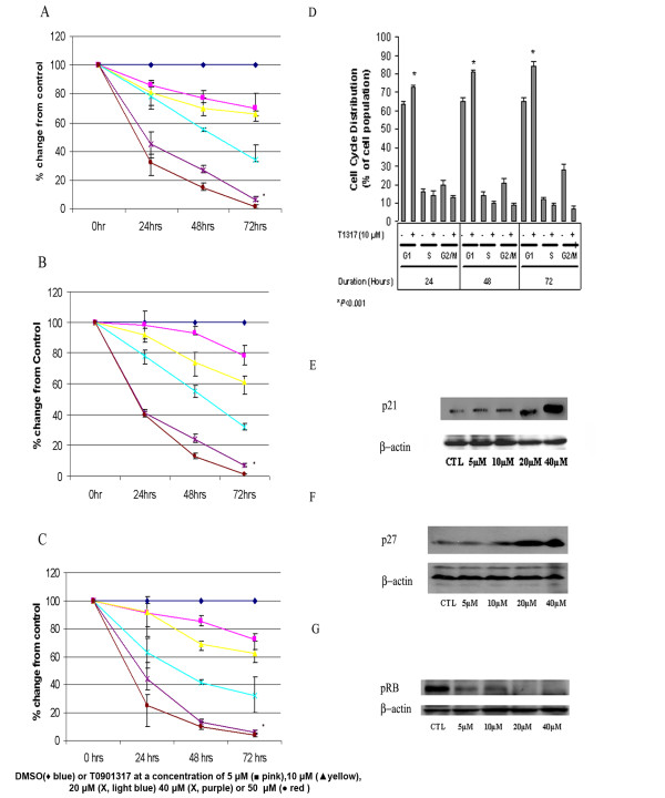
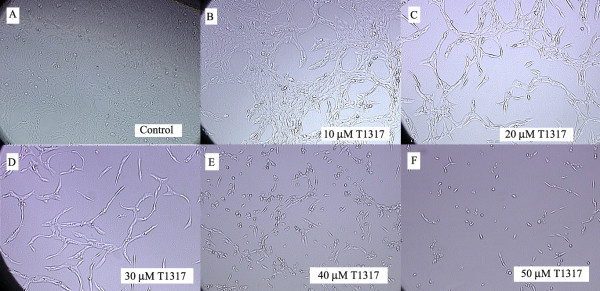
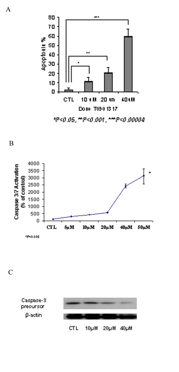
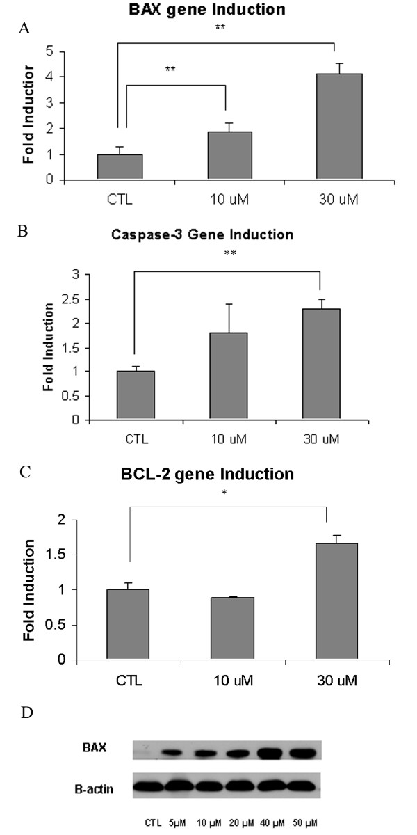
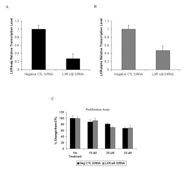
Results: T0901317 treatment resulted in a significant (P <0.001) inhibition of cell proliferation in a time- and dose-dependent manner in CaOV3, SKOV3 and A2780 cells. Western blot analysis demonstrated an induction of p21 and p27 with a concominant reduction in phospho-RB protein levels. Cell cycle analysis demonstrated a significant (P <0.001) arrest in the G1 cell cycle phase. Significant induction of Caspase-3 and BAX gene expression occurred with treatment. Induction of apoptosis was confirmed by significant (P < 0.001) elevation of caspase activity on FACS analysis, caspase-glo assay, BAX protein induction and decreased caspase 3 precursor protein expression on Western blot analysis. LXR alpha/beta knockdown experiments did not reverse the anti-proliferative and cytotoxic effects of T0901317.
Conclusions: The LXR agonist, T0901317, significantly suppresses cell proliferation and induces programmed cell death in a dose- and time-dependent manner. Our results indicate that T0901317 induces its anti-proliferative and cytotoxic effects via an LXR-independent mechanism.
Figures
Similar articles
-
miR-33 inhibition attenuates the effect of liver X receptor agonist T0901317 on expression of liver X receptor alpha in mice liver.ARYA Atheroscler. 2017 Nov;13(6):257-263. ARYA Atheroscler. 2017. PMID: 29643920 Free PMC article.
-
Activation of liver X receptors and retinoid X receptors induces growth arrest and apoptosis in insulin-secreting cells.Endocrinology. 2007 Apr;148(4):1843-9. doi: 10.1210/en.2006-1247. Epub 2006 Dec 28. Endocrinology. 2007. PMID: 17194744
-
Liver X receptor activation induces apoptosis of melanoma cell through caspase pathway.Cancer Cell Int. 2014 Feb 25;14(1):16. doi: 10.1186/1475-2867-14-16. Cancer Cell Int. 2014. PMID: 24564864 Free PMC article.
-
Liver X receptor agonist inhibits proliferation of ovarian carcinoma cells stimulated by oxidized low density lipoprotein.Gynecol Oncol. 2010 Jan;116(1):109-16. doi: 10.1016/j.ygyno.2009.09.034. Epub 2009 Oct 24. Gynecol Oncol. 2010. PMID: 19854496
-
LXR agonism improves TNF-α-induced endothelial dysfunction in the absence of its cholesterol-modulating effects.Atherosclerosis. 2014 Jan;232(1):1-9. doi: 10.1016/j.atherosclerosis.2013.10.001. Epub 2013 Oct 26. Atherosclerosis. 2014. PMID: 24401210
Cited by
-
RNA sequencing-based analysis of gallbladder cancer reveals the importance of the liver X receptor and lipid metabolism in gallbladder cancer.Oncotarget. 2016 Jun 7;7(23):35302-12. doi: 10.18632/oncotarget.9181. Oncotarget. 2016. PMID: 27167107 Free PMC article.
-
Cholesterol Metabolic Reprogramming in Cancer and Its Pharmacological Modulation as Therapeutic Strategy.Front Oncol. 2021 May 24;11:682911. doi: 10.3389/fonc.2021.682911. eCollection 2021. Front Oncol. 2021. PMID: 34109128 Free PMC article. Review.
-
Menin Maintains Cholesterol Content in Colorectal Cancer via Repression of LXR-Mediated Transcription.Cancers (Basel). 2023 Aug 16;15(16):4126. doi: 10.3390/cancers15164126. Cancers (Basel). 2023. PMID: 37627154 Free PMC article.
-
Role of Protein Regulators of Cholesterol Homeostasis in Immune Modulation and Cancer Pathophysiology.Endocrinology. 2025 Feb 27;166(4):bqaf031. doi: 10.1210/endocr/bqaf031. Endocrinology. 2025. PMID: 39951497 Review.
-
Lipid Metabolism as a Potential Target of Liver Cancer.J Hepatocell Carcinoma. 2024 Feb 14;11:327-346. doi: 10.2147/JHC.S450423. eCollection 2024. J Hepatocell Carcinoma. 2024. PMID: 38375401 Free PMC article. Review.
References
-
- Burg ME Van der, van Lent M, Buyse M. The effect of debulking surgery after induction chemotherapy on the prognosis in advanced epithelial ovarian cancer: Gynecological Cancer Cooperative Group of the European Organization for Research and Treatment of Cancer. N Engl J Med. 1995;10:629–34. doi: 10.1056/NEJM199503093321002. - DOI - PubMed
LinkOut - more resources
Full Text Sources
Other Literature Sources
Research Materials
