. 2010 May 26;11(1):64.
doi: 10.1186/1465-9921-11-64.

Inhibition of IFN-gamma-dependent antiviral airway epithelial defense by cigarette smoke

Affiliations

Inhibition of IFN-gamma-dependent antiviral airway epithelial defense by cigarette smoke

Modestos A Modestou et al. Respir Res. .

Abstract

Background: Although individuals exposed to cigarette smoke are more susceptible to respiratory infection, the effects of cigarette smoke on lung defense are incompletely understood. Because airway epithelial cell responses to type II interferon (IFN) are critical in regulation of defense against many respiratory viral infections, we hypothesized that cigarette smoke has inhibitory effects on IFN-gamma-dependent antiviral mechanisms in epithelial cells in the airway.

Methods: Primary human tracheobronchial epithelial cells were first treated with cigarette smoke extract (CSE) followed by exposure to both CSE and IFN-gamma. Epithelial cell cytotoxicity and IFN-gamma-induced signaling, gene expression, and antiviral effects against respiratory syncytial virus (RSV) were tested without and with CSE exposure.

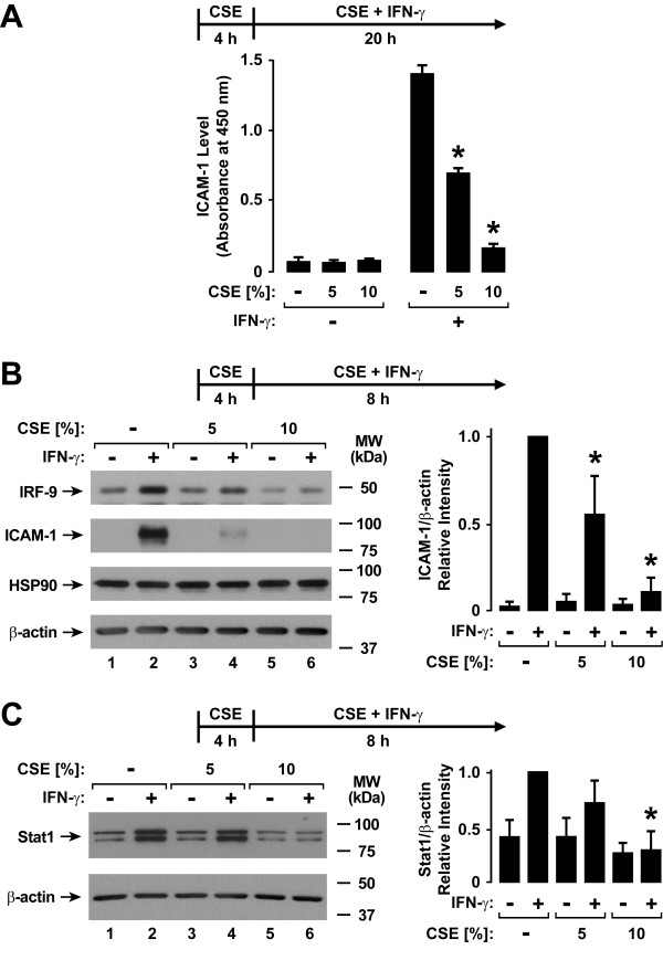
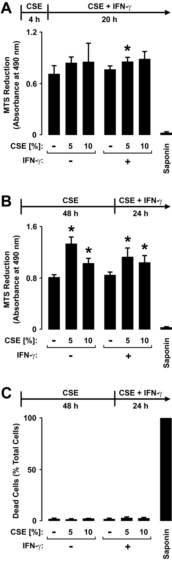
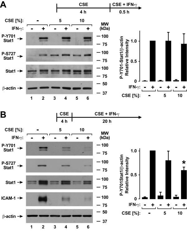
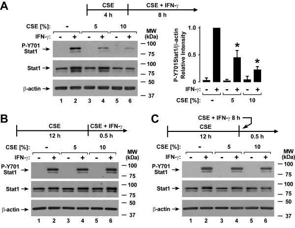
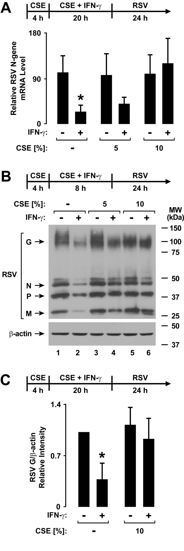
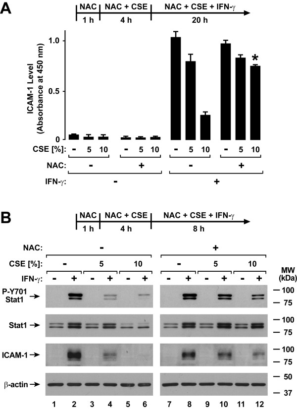
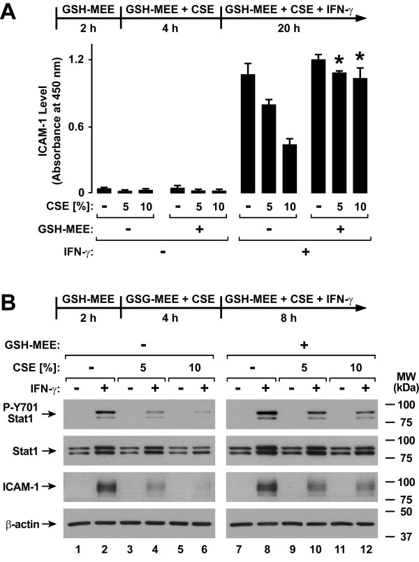
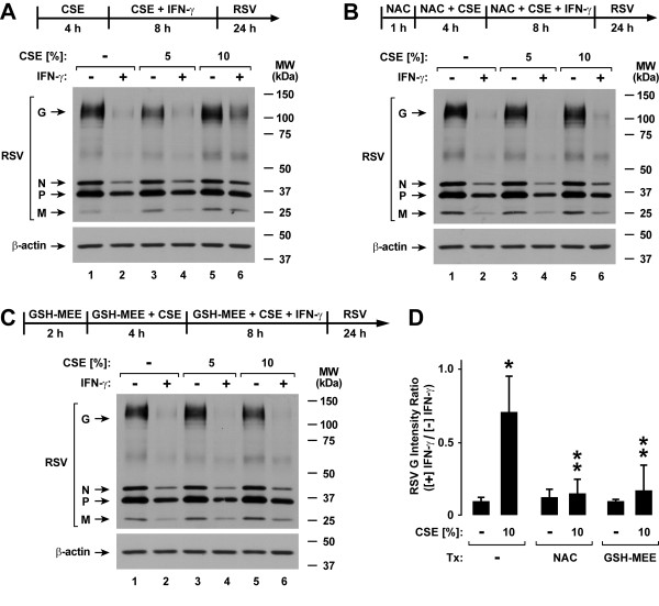
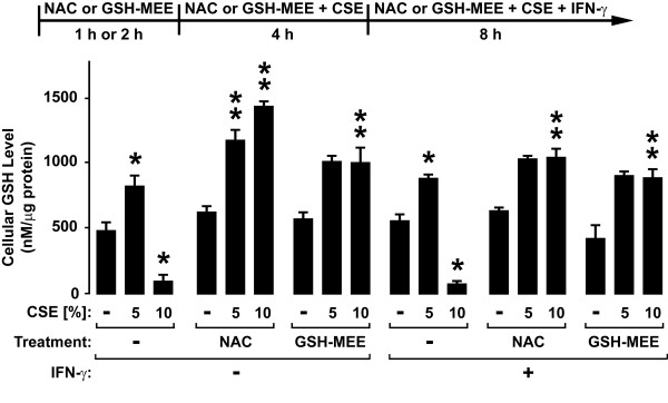
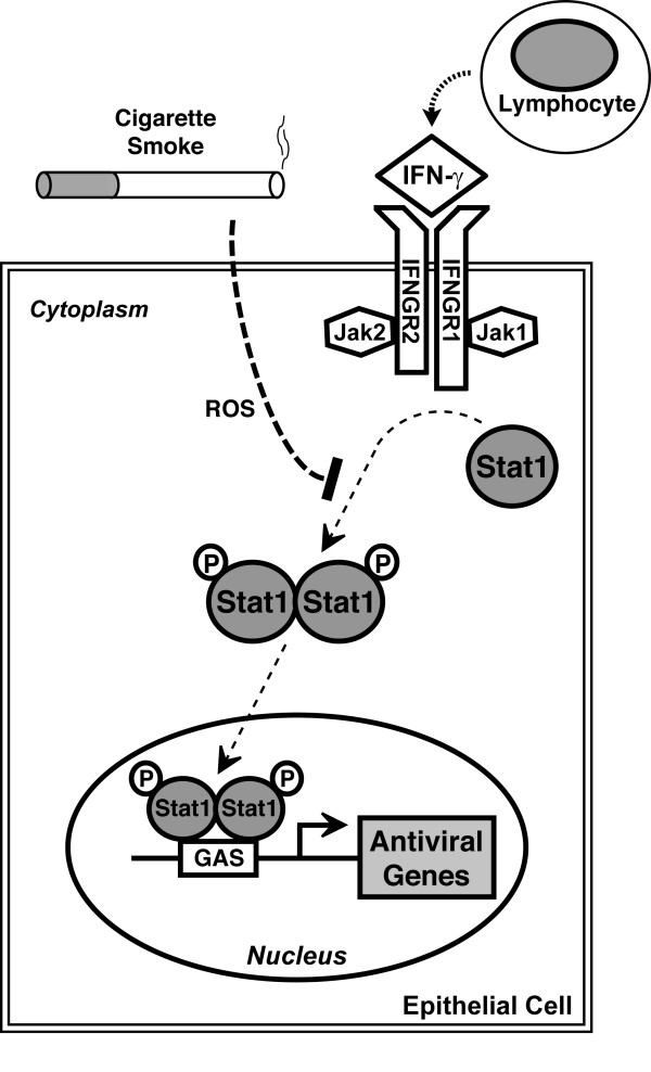
Results: CSE inhibited IFN-gamma-dependent gene expression in airway epithelial cells, and these effects were not due to cell loss or cytotoxicity. CSE markedly inhibited IFN-gamma-induced Stat1 phosphorylation, indicating that CSE altered type II interferon signal transduction and providing a mechanism for CSE effects. A period of CSE exposure combined with an interval of epithelial cell exposure to both CSE and IFN-gamma was required to inhibit IFN-gamma-induced cell signaling. CSE also decreased the inhibitory effect of IFN-gamma on RSV mRNA and protein expression, confirming effects on viral infection. CSE effects on IFN-gamma-induced Stat1 activation, antiviral protein expression, and inhibition of RSV infection were decreased by glutathione augmentation of epithelial cells using N-acetylcysteine or glutathione monoethyl ester, providing one strategy to alter cigarette smoke effects.

Conclusions: The results indicate that CSE inhibits the antiviral effects of IFN-gamma, thereby presenting one explanation for increased susceptibility to respiratory viral infection in individuals exposed to cigarette smoke.


Figures

Figure 1
Figure 1
Cigarette smoke extract decreases type II interferon-induced protein expression. A: ICAM-1 cell surface protein levels were determined using an enzyme-linked immunoassay with hTBE cell monolayers that were first treated with media without or with CSE at the indicated concentration for 4 hours. Cells were then incubated for 20 hours in media containing the same CSE concentration without or with IFN-γ. Values are expressed as mean ± S.D. (n = 3 replicates), and a significant difference (p < 0.05) in IFN-γ-induced levels between cells treated versus not treated with CSE is indicated by an asterisk. B: IRF-9, ICAM-1, HSP90, and β-actin protein levels were assessed using immunoblot analysis of extracts from hTBE cells that were first treated with media without or with CSE at the indicated concentration for 4 hours, followed by incubation for 8 hours in media containing the same CSE concentration without or with IFN-γ. C: Total Stat1 and β-actin protein levels were assessed using immunoblot analysis of extracts from hTBE cells that were first treated with media without or with CSE at the indicated concentration for 4 hours, followed by incubation for 8 hours in media containing the same CSE concentration without or with IFN-γ. In B and C, protein levels were quantified using band densitometry of immunoblot analyses from separate experiments with the value for cells not exposed to CSE but treated with IFN-γ set at 1 in each experiment. Values are expressed as mean ± S.D. (n = 4 experiments) in the bar graph adjacent to a representative immunoblot analysis, and a significant difference (p < 0.05) in IFN-γ-induced levels between cells treated versus not treated with CSE is indicated by an asterisk.
Figure 2
Figure 2
Cigarette smoke extract causes minimal airway epithelial cell cytotoxicity. A: Mitochondrial activity was quantified using an MTS-based assay with hTBE cells that were first treated with media without or with CSE at the indicated concentration for 4 hours, followed by incubation for 20 hours in media containing the same CSE concentration without or with IFN-γ. B: Mitochondrial activity was quantified using an MTS-based assay with hTBE cells that were first treated with media without or with CSE at the indicated concentration for 48 hours, followed by incubation for 24 hours in media containing the same CSE concentration without or with IFN-γ. In A and B, values are expressed as mean ± S.D. (n = 3 replicates). C: Dead and live hTBE cell numbers were quantified by detection of plasma membrane permeability to ethidium homodimers in dead cells and intracellular esterase activity in live cells. Cell monolayers were first treated with media without or with CSE at the indicated concentration for 48 hours, followed by incubation for 24 hours in media containing the same CSE concentration without or with IFN-γ. Values were calculated as dead cells/total cells and each condition represents the mean ± S.D. for 4 random low power fields (500-750 cells/field) from duplicate samples. In A-C, a significant difference (p < 0.05) in uninduced or IFN-γ-induced levels between cells treated versus not treated with CSE is indicated by an asterisk.
Figure 3
Figure 3
Cigarette smoke extract inhibits type II interferon-induced Stat1 activation. A: Tyrosine-701 and serine-727 phosphorylated and total Stat1, and β-actin levels were assessed using immunoblot analysis of extracts from hTBE cells that were first treated with media without or with CSE at the indicated concentrations for 4 hours, followed by incubation for 30 minutes in media containing the same CSE concentration without or with IFN-γ. B: Tyrosine-701 and serine-727 phosphorylated and total Stat1, ICAM-1, and β-actin levels were assessed using immunoblot analysis of extracts from hTBE cells that were first treated with media without or with CSE at the indicated concentrations for 4 hours, followed by incubation for 20 hours in media containing the same CSE concentration without or with IFN-γ. In A and B, protein levels were quantified using band densitometry of immunoblot analyses from separate experiments with the value for cells not exposed to CSE but treated with IFN-γ set at 1 in each experiment. Values are expressed as mean ± S.D. (n = 3 experiments) in the bar graph adjacent to a representative immunoblot analysis, and a significant difference (p < 0.05) in IFN-γ-induced levels between cells treated versus not treated with CSE is indicated by an asterisk.
Figure 4
Figure 4
Cigarette smoke extract has a delayed effect on epithelial cell Stat1 activation. A: Tyrosine-701 phosphorylated and total Stat1, and β-actin levels were assessed using immunoblot analysis of extracts from hTBE cells that were first treated with media without or with CSE at the indicated concentrations for 4 hours, followed by incubation for 8 hours in media containing the same CSE concentration without or with IFN-γ. Protein levels were quantified using band densitometry of immunoblot analyses from separate experiments with the value for cells not exposed to CSE but treated with IFN-γ set at 1 in each experiment. Values are expressed as mean ± S.D. (n = 3 experiments) in the bar graph adjacent to a representative immunoblot analysis, and a significant difference (p < 0.05) in IFN-γ-induced levels between cells treated versus not treated with CSE is indicated by an asterisk. B: Tyrosine-701 phosphorylated and total Stat1, and β-actin levels were assessed using immunoblot analysis of extracts from hTBE cells that were first treated with media without or with CSE at the indicated concentrations for 12 hours, followed by incubation for 30 minutes in media containing the same CSE concentration without or with IFN-γ. C: Tyrosine-701 phosphorylated and total Stat1, and β-actin levels were assessed using immunoblot analysis of extracts from hTBE cells that were first treated with media without or with CSE at the indicated concentrations for 12 hours, followed by incubation for 30 minutes in media without or with IFN-γ that had been preincubated without or with the same CSE concentration alone for 8 hours.
Figure 5
Figure 5
Cigarette smoke extract decreases type II interferon-dependent antiviral defense. A: RSV N-gene mRNA levels were determined using realtime RT-PCR analysis of total RNA from hTBE cells that were first treated with media without or with CSE at the indicated concentration for 4 hours, followed by incubation for 20 hours in media containing the same CSE concentration without or with IFN-γ. Cells were then infected with RSV for 24 hours. Values are expressed as mean normalized N-gene mRNA level compared to control HPRT mRNA level (n = 3-4 experiments), and a significant difference (p < 0.05) in untreated or CSE-treated levels between cells uninduced versus induced by IFN-γ is indicated by an asterisk. B: RSV and β-actin protein levels were assessed using immunoblot analysis of extracts from hTBE cells that were first treated with media without or with CSE at the indicated concentration for 4 hours, followed by incubation for 8 hours in media containing the same CSE concentration without or with IFN-γ. Cells were then infected with RSV for 24 hours. C: Protein levels were quantified using band densitometry of immunoblot analyses from separate experiments performed as outlined for B. The band intensity of RSV G/β-actin was set at 1 in each experiment for cells not exposed to CSE or IFN-γ and other conditions were compared relative to this value. Values are expressed as mean ± S.D. (n = 4 experiments), and a significant difference (p < 0.05) in untreated or CSE-treated levels between cells uninduced versus induced by IFN-γ is indicated by an asterisk.
Figure 6
Figure 6
N-acetylcysteine inhibits cigarette smoke effects on type II interferon-induced responses. A: ICAM-1 cell surface protein levels were determined using an enzyme-linked immunoassay with hTBE cells that were first treated in media without or with NAC for 1 hour. Cells were then incubated in media containing the antioxidant without or with CSE at the indicated concentrations for 4 hours, followed by incubation for 20 hours with the same compounds without or with IFN-γ. Values are expressed as mean ± S.D. (n = 3 replicates) and a significant difference (p < 0.05) in comparable CSE-treated levels between cells not incubated versus incubated with NAC is indicated by an asterisk. B: Tyrosine-701 phosphorylated and total Stat1, ICAM-1, and β-actin protein levels were assessed using immunoblot analysis of extracts from hTBE cells that were first treated in media without or with NAC for 1 hour. Cells were then incubated in media containing the antioxidant without or with CSE at the indicated concentrations for 4 hours, followed by incubation for 8 hours with the same compounds without or with IFN-γ.
Figure 7
Figure 7
GSH monoethyl ester inhibits cigarette smoke effects on type II interferon-induced responses. A: ICAM-1 cell surface protein levels were determined using an enzyme-linked immunoassay with hTBE cells that were first treated in media without or with GSH-MEE for 2 hours. Cells were then incubated in media containing the antioxidant without or with CSE at the indicated concentrations for 4 hours, followed by incubation for 20 hours with the same compounds without or with IFN-γ. Values are expressed as mean ± S.D. (n = 3 replicates) and a significant difference (p < 0.01) in comparable CSE-treated levels between cells not incubated versus incubated with GSH-MEE is indicated by an asterisk. B: Tyrosine-701 phosphorylated and total Stat1, ICAM-1, and β-actin protein levels were assessed using immunoblot analysis of extracts from hTBE cells that were first treated in media without or with GSH-MEE for 2 hours. Cells were then incubated in media containing the antioxidant without or with CSE at the indicated concentrations for 4 hours, followed by incubation for 8 hours with the same compounds without or with IFN-γ.
Figure 8
Figure 8
N-acetylcysteine and GSH monoethyl ester inhibit cigarette smoke effects on antiviral defense. RSV and β-actin protein levels were assessed using immunoblot analysis of extracts from hTBE cells that were first treated in media without (A) or with NAC for 1 hour (B) or GSH monoethyl ester for 2 hours (C). Cells were then incubated in media containing the same antioxidant without or with CSE at the indicated concentration for 4 hours, followed by incubation for 8 hours with the same compounds without or with IFN-γ. Cells were then infected with RSV for 24 hours. D: Protein levels were quantified using band densitometry of immunoblot analyses from separate experiments performed as outlined for A-C. The band intensities of RSV G/β-actin were compared between conditions by calculating ratios for each comparable condition in which cells were versus were not exposed to IFN-γ. Values are expressed as mean ± S.D. (n = 3 experiments), and a significant difference (p < 0.05) in levels between comparable antioxidant incubation conditions for cells without versus with CSE treatment is indicated by one asterisk, and in CSE-treated levels between cells not incubated versus incubated with NAC or GSH-MEE is indicated by two asterisks.
Figure 9
Figure 9
Cigarette smoke extract alters epithelial cell glutathione levels. Glutathione levels were determined using a luminescence-based assay with hTBE cells that were first treated in media without or with NAC for 1 hour or GSH-MEE for 2 hours. Cells were then incubated in media containing the antioxidant without or with CSE at the indicated concentration for 4 hours, followed by incubation for 8 hours with the same compounds without or with IFN-γ. Values are expressed as mean ± S.D. (n = 3 replicates), and a significant difference (p < 0.05) in uninduced or IFN-γ-induced levels between cells treated versus not treated with CSE but not exposed to antioxidant is indicated by one asterisk, and in comparable CSE-treated levels between cells not incubated versus incubated with NAC or GSH-MEE under uninduced or IFN-γ-induced conditions is indicated by two asterisks.
Figure 10
Figure 10
Model for cigarette smoke effects on type II interferon signal transduction. Cigarette smoke inhibits type II interferon-dependent gene expression by decreasing Stat1 phosphorylation. A portion of this effect is mediated by reactive oxygen species (ROS). Decreased antiviral gene expression decreases epithelial cell responses to IFN-γ that inhibit viral infection.

Similar articles

Cited by

References

    1. Gürkan F, Kiral A, Dagli E, Karakoç F. The effect of passive smoking on the development of respiratory syncytial virus bronchiolitis. Eur J Epidemiol. 2000;16:465–468. doi: 10.1023/A:1007658411953. - DOI - PubMed
    1. Bradley JP, Bacharier LB, Bonfiglio J, Schechtman KB, Strunk R, Storch G, Castro M. Severity of respiratory syncytial virus bronchiolitis is affected by cigarette smoke exposure and atopy. Pediatrics. 2005;115:e7–e14. - PubMed
    1. U.S. Department of Health and Human Services. The health consequences of involuntary exposure to tobacco smoke: a report of the Surgeon General. Department of Health and Human Services, Centers for Disease Control and Prevention, Coordinating Center for Health Promotion, National Center for Chronic Disease Prevention and Health Promotion, Office on Smoking and Health, Atlanta, GA; 2006.
    1. Benseñor IM, Cook NR, Lee IM, Chown MJ, Hennekens CH, Buring JE, Manson JE. Active and passive smoking and risk of colds in women. Ann Epidemiol. 2001;11:225–231. doi: 10.1016/S1047-2797(00)00214-3. - DOI - PubMed
    1. Blake GH, Abell TD, Stanley WG. Cigarette smoking and upper respiratory infection among recruits in basic combat training. Ann Intern Med. 1988;109:198–202. - PubMed

Publication types

MeSH terms