. 2010 Jul 23;285(30):23486-95.
doi: 10.1074/jbc.M110.142703. Epub 2010 May 26.

Complement regulator Factor H mediates a two-step uptake of Streptococcus pneumoniae by human cells

Affiliations

Complement regulator Factor H mediates a two-step uptake of Streptococcus pneumoniae by human cells

Vaibhav Agarwal et al. J Biol Chem. .

Abstract

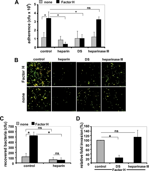
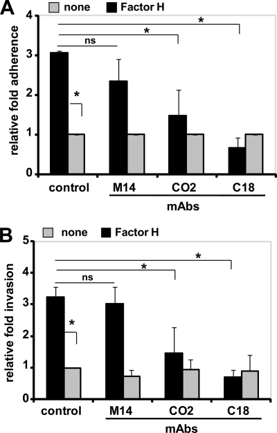
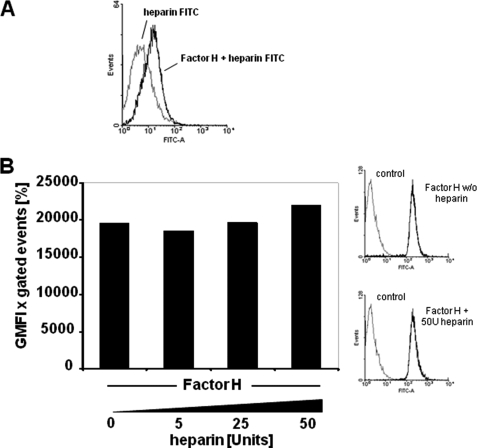
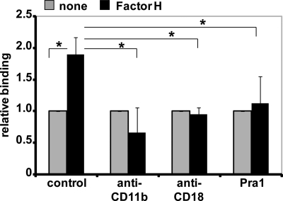
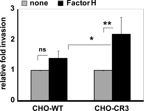
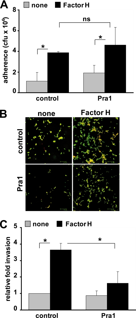
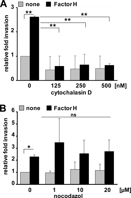
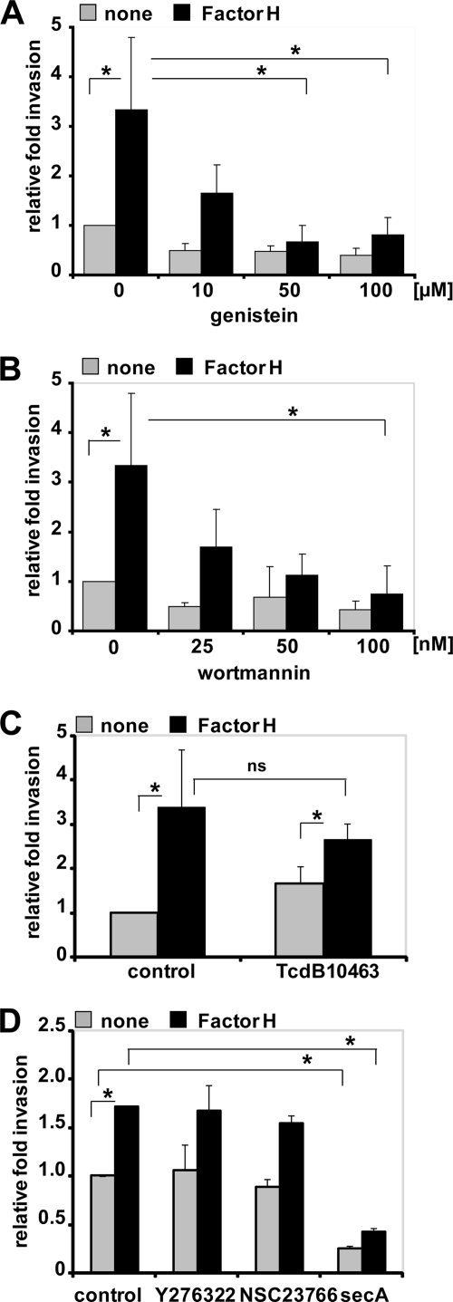
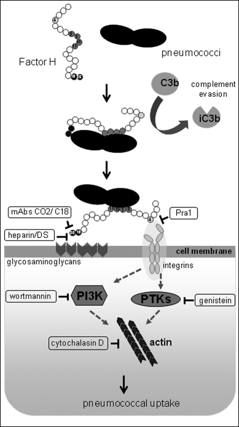
Streptococcus pneumoniae, a human pathogen, recruits complement regulator factor H to its bacterial cell surface. The bacterial PspC protein binds Factor H via short consensus repeats (SCR) 8-11 and SCR19-20. In this study, we define how bacterially bound Factor H promotes pneumococcal adherence to and uptake by epithelial cells or human polymorphonuclear leukocytes (PMNs) via a two-step process. First, pneumococcal adherence to epithelial cells was significantly reduced by heparin and dermatan sulfate. However, none of the glycosaminoglycans affected binding of Factor H to pneumococci. Adherence of pneumococci to human epithelial cells was inhibited by monoclonal antibodies recognizing SCR19-20 of Factor H suggesting that the C-terminal glycosaminoglycan-binding region of Factor H mediates the contact between pneumococci and human cells. Blocking of the integrin CR3 receptor, i.e. CD11b and CD18, of PMNs or CR3-expressing epithelial cells reduced significantly the interaction of pneumococci with both cell types. Similarly, an additional CR3 ligand, Pra1, derived from Candida albicans, blocked the interaction of pneumococci with PMNs. Strikingly, Pra1 inhibited also pneumococcal uptake by lung epithelial cells but not adherence. In addition, invasion of Factor H-coated pneumococci required the dynamics of host-cell actin microfilaments and was affected by inhibitors of protein-tyrosine kinases and phosphatidylinositol 3-kinase. In conclusion, pneumococcal entry into host cells via Factor H is based on a two-step mechanism. The first and initial contact of Factor H-coated pneumococci is mediated by glycosaminoglycans expressed on the surface of human cells, and the second step, pneumococcal uptake, is integrin-mediated and depends on host signaling molecules such as phosphatidylinositol 3-kinase.


Figures

FIGURE 1.
FIGURE 1.
Glycosaminoglycans inhibit Factor H-mediated pneumococcal adhesion to human lung epithelial cells. A, adherence of pneumococci via Factor H to lung epithelial A549 cells in the absence (control) or the presence of heparin (50 units/ml), dermatan sulfate (100 μg/ml), or after pretreatment with heparinase III (10 milliunits/ml) was estimated by quantifying the colony-forming units (cfu) per well obtained from plating onto blood agar plates. The infection assays were conducted with or without (none) pretreatment of pneumococci with Factor H. *, p < 0.02. B, immunofluorescence microscopy of Factor H-mediated pneumococcal adhesion after treatment of host cells with glycosaminoglycans or heparinase III. Adherent bacteria appear green/yellow (Alexa Fluor 488/568), and intracellular bacteria were stained red (Alexa Fluor 568). C and D, pneumococcal invasion into epithelial cells via Factor H is diminished in the presence of glycosaminoglycans. Pneumococcal invasion was determined after conducting infection assays in the absence (control) or presence of glycosaminoglycans by employing the antibiotic protection assay. *, p < 0.001 relative to infections carried out with Factor H but in the absence of inhibitor; ns, not significant.
FIGURE 2.
FIGURE 2.
C-terminal SCR19–20 of Factor H is the key co-factor in Factor H-mediated pneumococcal adherence to and invasion into host cells. A, adherence of Factor H-coated pneumococci to A549 cells was determined in the presence of mAbs M14, CO2, or C18 (each 2 μg ml−1 per well) or absence of a mAb (control). The antibodies bind to the middle region (M14), SCR19 (CO2), or SCR19–20 (C18) of Factor H. Results are shown as relative adherence of Factor H-bound pneumococci compared with untreated pneumococci. *, p < 0.02; ns, not significant. B, invasion and intracellular survival of Factor H-coated pneumococci in the presence of monoclonal antibodies M14, CO2, or C18 (each 2 μg ml−1) were determined by the antibiotic protection assay. Results show the relative invasion of pneumococci into A549 cells compared with Factor H-treated bacteria and in the absence (control) of monoclonal antibodies. *, p < 0.02; ns, not significant.
FIGURE 3.
FIGURE 3.
Effect of heparin on Factor H recruitment by pneumococci. A, binding of FITC-heparin (2 μg) to pneumococci or to Factor H-pretreated pneumococci was determined by flow cytometry. The histogram shows the log fluorescence intensity (FITC-A) on the x axis, and the y axis shows the number of events. B, binding of heparin-pretreated Factor H to pneumococci. The effect of heparin on pneumococcal recruitment of Factor H was analyzed by flow cytometry after preincubation of 2 μg of purified Factor H with the indicated amounts of heparin per reaction. Bacterial bound Factor H was determined by flow cytometry, and results were expressed as mean fluorescence intensity multiplied with the percentage of FITC-labeled bacteria. The graph shows a representative experiment. The results are also represented as histograms, where the x axis represents fluorescence of pneumococcus-associated Factor H, in the absence (w/o) or presence of 50 units of heparin, on a log10 scale, and the y axis represents the number of events.
FIGURE 4.
FIGURE 4.
Factor H promotes pneumococcal binding to PMNs via integrin CR3. Pneumococci were incubated with PMNs for 30 min, in the absence (control) or presence of anti-CD11b (2 μg), anti-CD18 (2 μg), or integrin CR3 (CD11b/CD18) ligand Pra1 (2 μg). Pneumococcal binding to PMNs was investigated in the absence (none) or presence of bacteria-bound Factor H by flow cytometry. The results were calculated (mean fluorescence intensity × percentage of gated positive events), and the data show the relative binding ratios compared with Factor H untreated pneumococci. *, p < 0.02.
FIGURE 5.
FIGURE 5.
Integrin CR3 (CD11b/CD18) promotes invasion of Factor H-coated pneumococci into epithelial cells. Invasion and intracellular survival of pneumococci in CHO-WT (CHO-K1 cells) and CHO-CR3 cells were determined by the antibiotic protection assay. The results are shown relative to infections conducted with untreated pneumococci. *, p < 0.05; **, p < 0.01; ns, not significant.
FIGURE 6.
FIGURE 6.
Factor H-mediated pneumococcal internalization by epithelial cells is inhibited by the integrin-binding protein Pra1. A, pneumococcal adherence to A549 cells was determined in the absence (control) or presence of integrin CR3 ligand Pra1 (2 μg ml−1). The infection assays were conducted with or without (none) pretreatment of pneumococci with Factor H. *, p < 0.03; ns, not significant, relative to infections conducted in the absence of Factor H. B, immunofluorescence microscopy of pneumococcal adherence via Factor H to A549 cells in the absence (control) or presence of Pra1. C, invasion and intracellular survival of pneumococci in A549 cells were monitored in the absence (control) or the presence of Pra1 (2 μg ml−1) by the antibiotic protection assay. The results are shown relative to infections conducted in the absence of Pra1 and Factor H. *, p < 0.02.
FIGURE 7.
FIGURE 7.
Factor H-mediated pneumococcal ingestion by epithelial cells requires actin cytoskeleton dynamics. A and B, pneumococcal invasion into A549 was determined in the presence of cytochalasin D (A) or nocodazole (B) by the antibiotic protection assay as described previously (47). Pneumococcal invasion indicates the relative number of intracellular bacteria (Factor H pretreated or untreated (none)) in the presence of an inhibitor compared with pneumococcal invasion in the absence of an inhibitor. *, p < 0.05; **, p < 0.001; ns, not significant.
FIGURE 8.
FIGURE 8.
Protein-tyrosine kinase activities and PI3K but not small Rho family GTPases are essential for Factor H-mediated pneumococcal ingestion by epithelial cells. The number of invasive pneumococci was determined in the presence of genistein (A), which is a phosphotyrosine kinase inhibitor, PI3K inhibitor wortmannin (B), C. difficile toxin B, TcdB10463 (30 ng ml−1) (C), or specific individual inhibitors of Rho family GTPases such as Y27632 (50 μm), Rac1 inhibitor NSC23766 (50 μm), or Cdc42 inhibitor secramine A (10 μm) (D) by employing the antibiotic protection assay. Shown is the relative invasion of Factor H pretreated or untreated (none) pneumococci in the presence of an inhibitor compared with pneumococcal invasion in the absence of an inhibitor. *, p < 0.05; ns, not significant, relative to infections in the presence of Factor H but absence of inhibitors.
FIGURE 9.
FIGURE 9.
Schematic model of the two-step Factor H-integrin complex-mediated pneumococcal epithelial cell invasion mechanism. PTK, protein-tyrosine kinase.

References

    1. Austrian R. (1986) J. Antimicrob. Chemother. 18, Suppl. A, 35–45 - PubMed
    1. Cartwright K. (2002) Eur. J. Pediatr. 161, 188–195 - PubMed
    1. Hammerschmidt S. (2006) Curr. Opin. Microbiol. 9, 12–20 - PubMed
    1. Musher D. M. (1992) Clin. Infect. Dis. 14, 801–807 - PubMed
    1. Bergmann S., Hammerschmidt S. (2006) Microbiology 152, 295–303 - PubMed

Publication types

MeSH terms