Comparative Study
. 2010 Aug 6;7 Suppl 4(Suppl 4):S403-10.
doi: 10.1098/rsif.2010.0134.focus. Epub 2010 May 26.

Needle-shaped polymeric particles induce transient disruption of cell membranes

Affiliations
Comparative Study

Needle-shaped polymeric particles induce transient disruption of cell membranes

Nishit Doshi et al. J R Soc Interface. .

Abstract

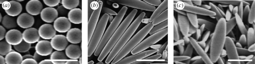
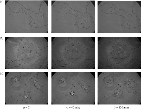
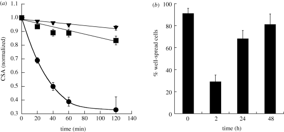
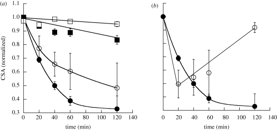
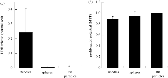
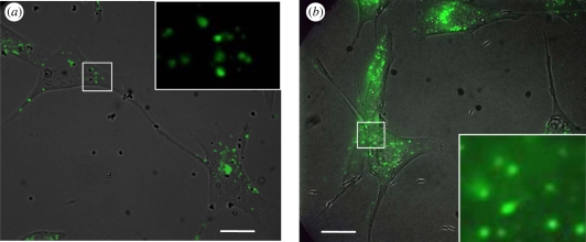
Nano- and microparticles of various shapes have recently been introduced for various drug-delivery applications. Shape of particles has been shown to have an impact on various processes including circulation, vascular adhesion and phagocytosis. Here, we assess the role of particle geometry and surface chemistry in their interactions with cell membranes. Using representative particles of different shape (spheres, elongated and flat particles), size (500 nm-1 microm) and surface chemistry (positively and negatively charged), we evaluated the response of endothelial cells to particles. While spherical and elliptical disc-shaped particles did not have an impact on cell spreading and motility, needle-shaped particles induced significant changes in the same. Further studies revealed that needle-shaped particles induced disruption of cell membranes as indicated by the release of lactate dehydrogenase and uptake of extracellular calcein. The effect of needle-shaped particles on cells was transient and was reversed over a time period of 1-48 h depending on particle parameters.


Figures

Figure 1.
Figure 1.
Scanning electron micrographs of particles of different geometry used for the study. (a) Spheres, (b) needles and (c) elliptical discs. Spheres are 1 µm in diameter, whereas needles and elliptical discs are stretched from these spheres. Scale bars, 2 µm.
Figure 2.
Figure 2.
Time-lapse video microscopy images of interaction of particles of different geometry with endothelial cells at different time points: spheres (a), elliptical discs (b) and needles (c). The cells incubated with spheres and elliptical discs do not show significant morphological changes over time, whereas in the case of needles, the cells undergo contraction and re-spread over time. Cell boundaries have been drawn manually as a visual aid.
Figure 3.
Figure 3.
Kinetics of cellular contractions caused by particles of different geometry on endothelial cells. (a) Plot of normalized CSA for spheres (squares), elliptical discs (triangles) and needles (circles) against time. Spheres are 1 µm in diameter, whereas needles and elliptical discs are stretched from these spheres. The CSA for spheres and elliptical discs varies at most 15 per cent over a period of 2 h, whereas for needles, the CSA decreases rapidly by more than 65 per cent in 2 h. (b) Percentage of well-spread cells before and after the treatment with needles for 2, 24 and 48 h. The percentage of well-spread cells decreases drastically 2 h after incubation with needles; however, the cells recover to the baseline in approximately 48 h.
Figure 4.
Figure 4.
Effect of size and surface chemistry on the kinetics of cellular contractions caused by particles of different shape on endothelial cells. (a) Effect of size of particles on normalized CSA. Spheres of two different sizes of 1 µm (filled squares) and 0.5 µm (open squares) and corresponding needles (filled circles and open circles, respectively) stretched from these spheres were used. Large-sized particles showed more drastic effects on endothelial cells. (b) Effect of plain needles (filled circles) and positively charged needles (open circles) on normalized CSA. Positively charged particles result in rapid reduction of CSA and rapid recovery compared with plain needles. The lines are drawn to guide the eye.
Figure 5.
Figure 5.
Toxicological assays with particles of different geometry on endothelial cells. (a) LDH release from endothelial cells upon interaction with particles of different geometry. One micrometre spheres and needles stretched from these spheres were used for the assay. Needles showed significantly higher LDH release when compared with spheres (p < 0.01, n = 5). (b) Proliferation potential of endothelial cells upon incubation with particles of different geometry as determined by the MTT assay. One micrometre spheres and needles stretched from these spheres were used for the assay. There was no significant difference in the proliferation potential of spheres and needles (p > 0.05, n = 5) and it was close to negative control.
Figure 6.
Figure 6.
Calcein uptake by endothelial cells incubated with particles of different shapes. The figures show overlaid brightfield and fluorescent images. (a) 1 µm spheres. The punctuate fluorescent spots clearly indicate pinocytic uptake of calcein. (b) Needles stretched from 1 µm spheres. The diffuse fluorescence of calcein in the cell membrane indicates transmembrane uptake in this case. Scale bar, 20 µm. The insets show enlarged images of sections shown by white rectangles in the main images. The insets show only fluorescent images.

Similar articles

Cited by

References

    1. Alexis F., Pridgen E., Molnar L. K., Farokhzad O. C. 2008. Factors affecting the clearance and biodistribution of polymeric nanoparticles. Mol. Pharm. 5, 505–515. (10.1021/mp800051m) - DOI - PMC - PubMed
    1. Allen T. M., Cullis P. R. 2004. Drug delivery systems: entering the mainstream. Science 303, 1818–1822. (10.1126/science.1095833) - DOI - PubMed
    1. Beningo K. A., Wang Y. L. 2002. Fc-receptor-mediated phagocytosis is regulated by mechanical properties of the target. J. Cell Sci. 115, 849–856. - PubMed
    1. Brannon-Peppas L. 1995. Recent advances on the use of biodegradable microparticles and nanoparticles in controlled drug delivery. Int. J. Pharm. 116, 1–9. (10.1016/0378-5173(94)00324-X) - DOI
    1. Brigger I., Dubernet C., Couvreur P. 2002. Nanoparticles in cancer therapy and diagnosis. Adv. Drug Deliv. Rev. 54, 631–651. (10.1016/S0169-409X(02)00044-3) - DOI - PubMed

Publication types

LinkOut - more resources