. 2010 Aug;106(2):235-42.
doi: 10.1093/aob/mcq101. Epub 2010 May 26.

Hourglass cell development in the soybean seed coat

Affiliations

Hourglass cell development in the soybean seed coat

S S Miller et al. Ann Bot. 2010 Aug.

Abstract

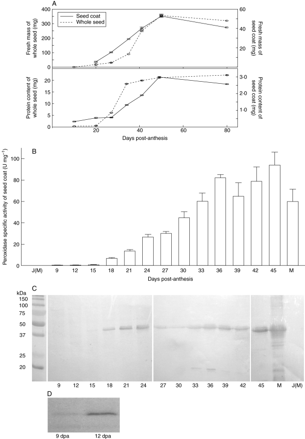
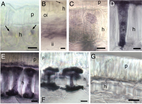
Background and aims: Hourglass cells (HGCs) are prominent cells in the soybean seed coat, and have potential use as 'phytofactories' to produce specific proteins of interest. Previous studies have shown that HGCs initiate differentiation at about 9 d post-anthesis (dpa), assuming their characteristic morphology by 18 dpa. This study aims to document the structural changes in HGCs during this critical period, and to relate these changes to the concurrent development of a specific soybean peroxidase (SBP) encoded by the Ep gene.

Methods: Pods were collected from plants at specific growth stages. Fresh material was processed for analysis of Ep peroxidase activity. Tissues were processed for scanning and transmission electron microscopy, as well as extracted for western blotting. A null variety lacking expression of Ep peroxidase was grown as a control.

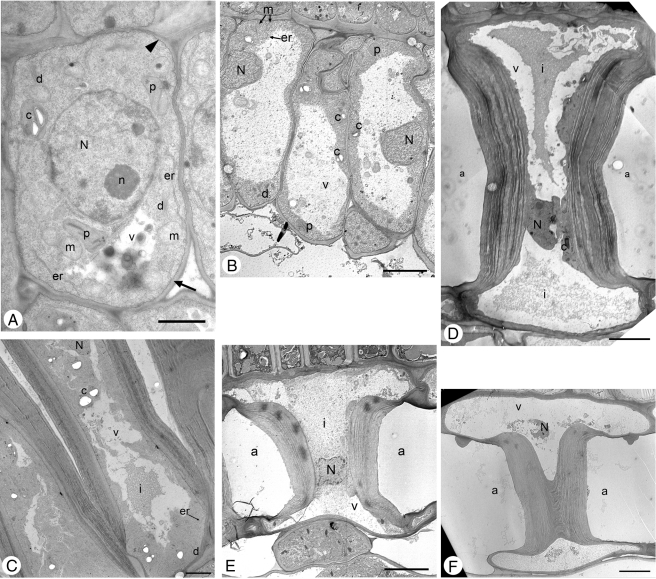
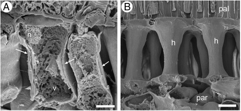
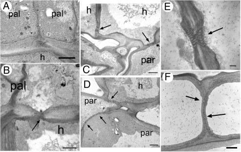
Key results and conclusions: At 9 dpa, HGCs are typical undifferentiated plant cells, but from 12-18 dpa they undergo rapid changes in their internal and external structure. By 18 dpa, they have assumed the characteristic hourglass shape with thick cell walls, intercellular air spaces and large central vacuoles. By 45 dpa, all organelles in HGCs have been degraded. Additional observations indicate that plasmodesmata connect all cell types. SBP activity and SBP protein are detectable in the HGC before they are fully differentiated (approx. 18 dpa). In very early stages, SBP activity appears localized in a vacuole as previously predicted. These results increase our understanding of the structure and development of the HGC and will be valuable for future studies aimed at protein targeting to components of the HGC endomembrane systems.


Figures

Fig. 1.
Fig. 1.
Transmission electron micrographs of soybean seed coat, ‘Maple Presto’: (A) at 9 dpa, the chloroplast contains two starch granules (arrow, cell wall; arrowhead, plasma membrane); (B) at 12 dpa, the chloroplast contains starch granules, the plasma membrane is visible inside cell walls but the tonoplast is not distinguishable; (C) at 15 dpa, the chloroplast contains starch granules but plastid itself is breaking down, the mitochondria are barely visible, the plasma membrane is visible inside the cell walls, although with some interruptions but the tonoplast is not distinguishable; (D) at 18 dpa, the nucleolus is visible inside the nucleus; (E) at 21 dpa; (F) at 45 dpa (glancing section). Abbreviations: a, air space between cells; c, chloroplast; d, dictyosome; er, endoplasmic reticulum; g, dictyosome; i, vacuolar inclusions; m, mitochondria; N, nucleus; n, nucleolus; p, proplastids; v, vacuole. Scale bars: (A) = 2 µm; (B–F) = 10 µm.
Fig. 2.
Fig. 2.
SEM images of soybean seed coat ‘Maple Presto’: (A) at 15 dpa (note multiple cell wall layers indicated by arrows); (B) at 18 dpa. Abbreviations: h, hourglass cell; n, nucleus; pal, palisade layer; par, parenchyma; v, vacuole. Scale bars: (A) = 5 µm; (B) = 20 µm.
Fig. 3.
Fig. 3.
TEM of soybean seed coat, ‘Maple Presto’, showing plasmodesmata connecting the different layers of the seed coat: (A) between palisade cells at 15 dpa; (B) between palisade and HGC at 15 dpa; (C) between HGCs, and between HGC and parenchyma at 15 dpa; (D) between HCG and parenchyma, and between parenchyma cells at 15 dpa; (E) between HCCs at the bottom of the cells at 30 dpa; (F) between HCGs at the bottom of the cells at 45 dpa. Abbreviations: n, nucleus; pal, palisade layer; par, parenchyma. Scale bars: (A) = 5 µm; (B) = 2 µm; (C, D) = 2·5 µm; (E) = 0·5 µm; (F) = 1·0 µm.
Fig. 4.
Fig. 4.
Seed coat development and the production of soybean peroxidase: (A) profile of soybean seed development in soybean ‘Harosoy 63’ (EpEp). Top: individual seed and seed coat fresh mass at different developmental stages, as indicated (n ≥ 12). Bottom: protein content per seed and seed coat at different developmental stages (n ≥ 4). The bars indicate s.e. (B) Mean peroxidase specific activity of seed coat extracts from the soybean ‘Harosoy 63’ (EpEp) at different developmental stages (n = 3). The epep ‘Jack’ was used as a control. M and J(M) are samples from mature seed of ‘Harosoy 63’ and ‘Jack’, respectively. Bars indicate s.e. (C) Developmental expression of soybean peroxidase as detected by immunoblot analysis of seed coat extracts from soybean ‘Harosoy 63’ (EpEp). The epep ‘Jack’ was used as a control. Samples containing 10 µg of extract from 9 to 45dpa were analysed. M and J(M) are samples from mature seed of ‘Harosoy 63’ and ‘Jack’, respectively. Similar results were obtained for three replicates. (D) Samples from seed coats of soybean ‘Harosoy 63’ (EpEp) taken at 9 and 12 dpa containing 60 µg of extract as detected by immunoblot analysis.
Fig. 5.
Fig. 5.
Peroxidase activity in the developing soybean seed coat as visualized by chloronaphthol staining: (A–F) unfixed sections (125 µm) of ‘Harosoy 63’ at 9 dpa (A, B), 10 dpa (C), 13 dpa (D), 15 dpa (E) and mature seed coats (F). Mature, unfixed seed coat from ‘Jack’ (epep) was used as a control (G). Arrows point to vesicles. Abbreviations: h, hourglass cells; p, palisade cells, ii, inner integument, oi, outer integument. Scale bars: (A–F) = 10 µm; (G) = 20 µm.

References

    1. Bradford MM. A rapid and sensitive method for the quantification of microgram quantities of protein utilizing the principle of protein-dye binding. Analytical Biochemistry. 1976;72:248–254. - PubMed
    1. Corner EJH. The leguminous seed. Phytomorphology. 1951;1:117–150.
    1. Gijzen M. A deletion mutation at the ep locus causes low seed coat peroxidase activity in soybean. The Plant Journal. 1997;12:991–998. - PubMed
    1. Gijzen M, van Huystee R, Buzzell RI. Soybean seed coat peroxidase. Plant Physiology. 1993;103:1061–1066. - PMC - PubMed
    1. Gijzen M, Miller SS, Bowman L-A, Batchelor AK, Boutilier K, Miki BLA. Localization of peroxidase mRNAs in soybean seeds by in situ hybridization. Plant Molecular Biology. 1999;41:57–63. - PubMed

Publication types

MeSH terms