. 2010 Aug;84(15):7793-802.
doi: 10.1128/JVI.02643-09. Epub 2010 May 26.

The capsid protein of Turnip crinkle virus overcomes two separate defense barriers to facilitate systemic movement of the virus in Arabidopsis

Affiliations

The capsid protein of Turnip crinkle virus overcomes two separate defense barriers to facilitate systemic movement of the virus in Arabidopsis

Mingxia Cao et al. J Virol. 2010 Aug.

Abstract

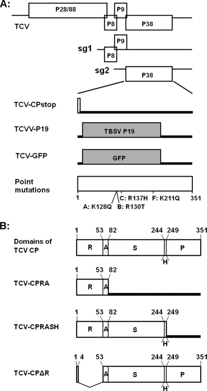
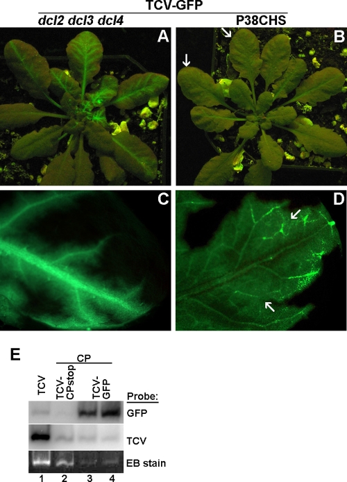
The capsid protein (CP) of Turnip crinkle virus (TCV) is a multifunctional protein needed for virus assembly, suppression of RNA silencing-based antiviral defense, and long-distance movement in infected plants. In this report, we have examined genetic requirements for the different functions of TCV CP and evaluated the interdependence of these functions. A series of TCV mutants containing alterations in the CP coding region were generated. These alterations range from single-amino-acid substitutions and domain truncations to knockouts of CP translation. The latter category also contained two constructs in which the CP coding region was replaced by either the cDNA of a silencing suppressor of a different virus or that of green fluorescent protein. These mutants were used to infect Arabidopsis plants with diminished antiviral silencing capability (dcl2 dcl3 dcl4 plants). There was a strong correlation between the ability of mutants to reach systemic leaves and the silencing suppressor activity of mutant CP. Virus particles were not essential for entry of the viral genome into vascular bundles in the inoculated leaves in the absence of antiviral silencing. However, virus particles were necessary for egress of the viral genome from the vasculature of systemic leaves. Our experiments demonstrate that TCV CP not only allows the viral genome to access the systemic movement channel through silencing suppression but also ensures its smooth egress by way of assembled virus particles. These results illustrate that efficient long-distance movement of TCV requires both functions afforded by the CP.


Figures

FIG. 1.
FIG. 1.
Schematic representation of the genome structure of TCV and its mutants. (A) Top three diagrams depict the genomic (g) and subgenomic (sg) RNAs of TCV, with all the open reading frames (ORFs) shown as open boxes. The bottom four diagrams show the CP region of mutants TCV-CPstop, TCVV-P19, and TCV-GFP and four single-amino-acid mutants (A, B, C, and F), with the open or shaded boxes denoting the expected translation products and the thick lines representing the RNA regions not expected to be translated. The exact amino acid changes in the single-amino-acid mutants are marked beneath the bottom diagram, together with their approximate positions. (B) Top diagram represents the full-length CP of TCV, with the sizes and the relative positions of the five structural domains shown. The numbers on the top are the positions of the first amino acids of the respective domains and the last amino acid of the whole CP. R, RNA-binding domain; A, arm; S, surface domain; H, hinge; P, protruding domain. The next three diagrams depict three mutants with truncated CPs. TCV-CPΔR contains an in-frame deletion, whereas both CPRA and CPRASH are mutants that prevent full CP translation with added stop codons.
FIG. 2.
FIG. 2.
Alignment of multiple CPs of viruses in the Tombusviridae family. The four basic amino acid residues conserved in carmoviruses but not in tombusviruses are highlighted by asterisks immediately underneath.
FIG. 3.
FIG. 3.
Mutations within TCV CP differentially affect its functions in silencing suppression, virion assembly, and viral systemic spread. (A) Evaluation of the silencing suppression capability of different TCV CP mutants using the agro-infiltration assay. TBSV P19 (leaf no. 4) was used as an additional positive control. The leaves were photographed at 5 days after infiltration. (B) Detection of GFP mRNA and siRNAs in the agro-infiltrated leaves with RNA blot hybridizations. (C) dsRNA-binding properties of CPA, CPB, CPC, and CPF mutants. Top, 5% nondenaturing polyacrylamide gel containing protein extracts preinoculated with 32P-labeled dsRNA. Bottom, 12% SDS-PAGE gel serving showing the relative concentrations of mutant proteins used in the binding assay. (D) Systemic symptoms on Col-0 and dcl2 dcl3 dcl4 plants infected with various TCV mutants. Mock, buffer inoculated. Photographs were taken at 18 dpi. (E) Close-up images of dcl2 dcl3 dcl4 plants not inoculated (top left), inoculated with TCV-CPstop (bottom left), and inoculated with CPC (bottom right). Top right: systemically infected leaves taken from these plants. A relatively healthy class II vein on CPC-infected leaves is highlighted with an arrow. (F) Viral RNA, virion, and CP accumulations in plants infected with various TCV mutants as determined with RNA blot and Western blot hybridizations. Total RNA, protein, and virions were extracted from pooled systemic leaves (see Materials and Methods) at 11 to 12 dpi. Note that in the bottom two panels, lanes 5 and 6 were dilutions of the lane 4 sample. The panel at the very bottom is the stained gel serving as the control for protein load, with the TCV CP band easily visible.
FIG. 4.
FIG. 4.
Relative accumulation levels of viral RNAs and CP (or its truncated forms) of different CP deletion mutants. (A) RNA blot hybridization showing the viral RNAs of TCV-RA, TCV-RASH, and TCV-ΔR mutants in both wild-type Col-0 and dcl2 dcl3 dcl4 plants at 12 dpi. (B) Western blot showing the accumulation of CP or its derivatives in the systemically infected leaves. The white arrows in the stained gel (bottom) mark the positions of CP variants.
FIG. 5.
FIG. 5.
Different patterns of systemic movement of TCV-GFP in dcl2 dcl3 dcl4 and P38CHS plants. (A and B) TCV-GFP-infected dcl2 dcl3 dcl4 (A) and P38CHS (B) plants, photographed at 18 dpi. (C) A systemic leaf of a TCV-GFP-infected dcl2 dcl3 dcl4 plant viewed under a fluorescent microscope. (D) A systemic leaf of a TCV-GFP-infected P38CHS plant viewed under a fluorescent microscope. (E) A virion gel and its RNA blots showing virus particles assembled in N. benthamiana leaves infiltrated with CP+TCV-GFP constructs.

References

    1. Carrington, J. C., T. J. Morris, P. G. Stockley, and S. C. Harrison. 1987. Structure and assembly of turnip crinkle virus. IV. Analysis of the coat protein gene and implications of the subunit primary structure. J. Mol. Biol. 194:265-276. - PubMed
    1. Chen, M.-H., and V. Citovsky. 2003. Systemic movement of a tobamovirus requires host cell pectin methylesterase. Plant J. 35:386-392. - PubMed
    1. Choi, C. W., F. Qu, T. Ren, X. Ye, and T. J. Morris. 2004. The RNA silencing suppressor function of Turnip crinkle virus coat protein cannot be attributed to its interaction with the Arabidopsis protein TIP. J. Gen. Virol. 85:3415-3420. - PubMed
    1. Cohen, Y., A. Gisel, and P. C. Zambryski. 2000. Cell-to-cell and systemic movement of recombinant green fluorescent protein-tagged turnip crinkle viruses. Virology 273:258-266. - PubMed
    1. Deleris, A., J. Gallego-Bartolome, J. Bao, K. D. Kasschau, J. C. Carrington, and O. Voinnet. 2006. Hierarchical action and inhibition of plant Dicer-like proteins in antiviral defense. Science 313:68-71. - PubMed

Publication types

LinkOut - more resources