Interleukin-22 (IL-22) production by pulmonary Natural Killer cells and the potential role of IL-22 during primary influenza virus infection
- PMID: 20504940
- PMCID: PMC2897635
- DOI: 10.1128/JVI.00187-10
Interleukin-22 (IL-22) production by pulmonary Natural Killer cells and the potential role of IL-22 during primary influenza virus infection
Abstract
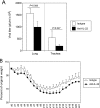
We set out to test the hypothesis that interleukin-22 (IL-22), a cytokine crucial for epithelial cell homeostasis and recovery from tissue injury, would be protective during influenza virus infection. Recent studies have identified phenotypically and functionally unique intestinal NK cells capable of producing the cytokine IL-22. Unlike gut NK cells that produce IL-22, the surface phenotypes of lung NK cells were similar to those of spleen NK cells and were characteristically mature. With mitogen stimulation, both single and double IL-22- and gamma interferon (IFN-gamma)-producing lung NK cells were detected. However, only the IL-22(+) IFN-gamma(-) lung NK subset was observed after stimulation with IL-23. IL-23 receptor (IL-23R) blocking dramatically inhibited IL-22 production, but not IFN-gamma production. Furthermore, we found that NK1.1(+) or CD27(-) lung NK cells were the primary sources of IL-22. After influenza virus infection, lung NK cells were quickly activated to produce both IFN-gamma and IL-22 and had increased cytotoxic potential. The level of IL-22 in the lung tissue declined shortly after infection, gradually returning to the baseline after virus clearance, although the IL-22 gene expression was maintained. Furthermore, depletion of NK cells with or without influenza virus infection reduced the protein level of IL-22 in the lung. Anti-IL-22 neutralization in vivo did not dramatically affect weight loss and survival after virus clearance. Unexpectedly, anti-IL-22-treated mice had reduced virus titers. Our data suggest that during primary respiratory viral infection, IL-22 seems to a play a marginal role for protection, indicating a differential requirement of this cytokine for bacterial and viral infections.
Figures


Similar articles
-
Type I interferon plays opposing roles in cytotoxicity and interferon-γ production by natural killer and CD8 T cells after influenza A virus infection in mice.J Innate Immun. 2014;6(4):456-66. doi: 10.1159/000356824. Epub 2014 Jan 10. J Innate Immun. 2014. PMID: 24435166 Free PMC article.
-
IL-22 from conventional NK cells is epithelial regenerative and inflammation protective during influenza infection.Mucosal Immunol. 2013 Jan;6(1):69-82. doi: 10.1038/mi.2012.49. Epub 2012 Jun 27. Mucosal Immunol. 2013. PMID: 22739232 Free PMC article.
-
Natural Killer Cells and Innate Interferon Gamma Participate in the Host Defense against Respiratory Vaccinia Virus Infection.J Virol. 2015 Oct 14;90(1):129-41. doi: 10.1128/JVI.01894-15. Print 2016 Jan 1. J Virol. 2015. PMID: 26468539 Free PMC article.
-
Role of interleukin-12 in primary influenza virus infection.J Virol. 1998 Jun;72(6):4825-31. doi: 10.1128/JVI.72.6.4825-4831.1998. J Virol. 1998. PMID: 9573248 Free PMC article.
-
IL-27 promotes NK cell effector functions via Maf-Nrf2 pathway during influenza infection.Sci Rep. 2019 Mar 21;9(1):4984. doi: 10.1038/s41598-019-41478-6. Sci Rep. 2019. PMID: 30899058 Free PMC article.
Cited by
-
IL-22: A key inflammatory mediator as a biomarker and potential therapeutic target for lung cancer.Heliyon. 2024 Aug 10;10(17):e35901. doi: 10.1016/j.heliyon.2024.e35901. eCollection 2024 Sep 15. Heliyon. 2024. PMID: 39263114 Free PMC article. Review.
-
Exposure to combustion derived particulate matter exacerbates influenza infection in neonatal mice by inhibiting IL22 production.Part Fibre Toxicol. 2021 Dec 14;18(1):43. doi: 10.1186/s12989-021-00438-7. Part Fibre Toxicol. 2021. PMID: 34906172 Free PMC article.
-
Neonatal hyperoxia leads to persistent alterations in NK responses to influenza A virus infection.Am J Physiol Lung Cell Mol Physiol. 2015 Jan 1;308(1):L76-85. doi: 10.1152/ajplung.00233.2014. Epub 2014 Nov 7. Am J Physiol Lung Cell Mol Physiol. 2015. PMID: 25381024 Free PMC article.
-
IL-22 Plays a Critical Role in Maintaining Epithelial Integrity During Pulmonary Infection.Front Immunol. 2020 Jun 9;11:1160. doi: 10.3389/fimmu.2020.01160. eCollection 2020. Front Immunol. 2020. PMID: 32582219 Free PMC article. Review.
-
Identification of Reference Genes in Chicken Intraepithelial Lymphocyte Natural Killer Cells Infected with Very-virulent Infectious Bursal Disease Virus.Sci Rep. 2020 May 22;10(1):8561. doi: 10.1038/s41598-020-65474-3. Sci Rep. 2020. PMID: 32444639 Free PMC article.
References
-
- Aktas, E., U. C. Kucuksezer, S. Bilgic, G. Erten, and G. Deniz. 2009. Relationship between CD107a expression and cytotoxic activity. Cell Immunol. 254:149-154. - PubMed
-
- Alter, G., J. M. Malenfant, and M. Altfeld. 2004. CD107a as a functional marker for the identification of natural killer cell activity. J. Immunol. Methods 294:15-22. - PubMed
-
- Anfossi, N., P. Andre, S. Guia, C. S. Falk, S. Roetynck, C. A. Stewart, V. Breso, C. Frassati, D. Reviron, D. Middleton, F. Romagne, S. Ugolini, and E. Vivier. 2006. Human NK cell education by inhibitory receptors for MHC class I. Immunity 25:331-342. - PubMed
-
- Aujla, S. J., Y. R. Chan, M. Zheng, M. Fei, D. J. Askew, D. A. Pociask, T. A. Reinhart, F. McAllister, J. Edeal, K. Gaus, S. Husain, J. L. Kreindler, P. J. Dubin, J. M. Pilewski, M. M. Myerburg, C. A. Mason, Y. Iwakura, and J. K. Kolls. 2008. IL-22 mediates mucosal host defense against Gram-negative bacterial pneumonia. Nat. Med. 14:275-281. - PMC - PubMed
-
- Aujla, S. J., and J. K. Kolls. 2009. IL-22: a critical mediator in mucosal host defense. J. Mol. Med. 87:451-454. - PubMed
Publication types
MeSH terms
Substances
Grants and funding
LinkOut - more resources
Full Text Sources
Other Literature Sources
Research Materials

