. 2010 Sep;88(3):495-505.
doi: 10.1189/jlb.1209822. Epub 2010 May 26.

Functional overlap but differential expression of CSF-1 and IL-34 in their CSF-1 receptor-mediated regulation of myeloid cells

Affiliations

Functional overlap but differential expression of CSF-1 and IL-34 in their CSF-1 receptor-mediated regulation of myeloid cells

Suwen Wei et al. J Leukoc Biol. 2010 Sep.

Abstract

CSF-1 is broadly expressed and regulates macrophage and osteoclast development. The action and expression of IL-34, a novel CSF-1R ligand, were investigated in the mouse. As expected, huIL-34 stimulated macrophage proliferation via the huCSF-1R, equivalently to huCSF-1, but was much less active at stimulating mouse macrophage proliferation than huCSF-1. Like muCSF-1, muIL-34 and a muIL-34 isoform lacking Q81 stimulated mouse macrophage proliferation, CSF-1R tyrosine phosphorylation, and signaling and synergized with other cytokines to generate macrophages and osteoclasts from cultured progenitors. However, they respectively possessed twofold and fivefold lower affinities for the CSF-1R and correspondingly, lower activities than muCSF-1. Furthermore, muIL-34, when transgenically expressed in a CSF-1-dependent manner in vivo, rescued the bone, osteoclast, tissue macrophage, and fertility defects of Csf1(op)/(op) mice, suggesting similar regulation of CSF-1R-expressing cells by IL-34 and CSF-1. Whole-mount IL34 in situ hybridization and CSF-1 reporter expression revealed that IL34 mRNA was strongly expressed in the embryonic brain at E11.5, prior to the expression of Csf1 mRNA. QRT-PCR revealed that compared with Csf1 mRNA, IL34 mRNA levels were lower in pregnant uterus and in cultured osteoblasts, higher in most regions of the brain and heart, and not compensatorily increased in Csf1(op/op) mouse tissues. Thus, the different spatiotemporal expression of IL-34 and CSF-1 allows for complementary activation of the CSF-1R in developing and adult tissues.


Figures
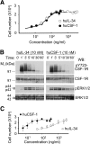
Figure 1.
Figure 1.
huCSF-1 and huIL-34 stimulated macrophage proliferation and signaling via the CSF-1R. (A) Proliferation dose-response curves of M−/− mouse macrophages expressing the huCSF-1R (M−/−.huCSF-1R macrophages; triplicate cultures±sem). (B) Kinetics of CSF-1R tyrosine 723 phosphorylation (pY723) and ERK1/2 phosphorylation (pERK1/2) of M−/−.huCSF-1R macrophages in response to huIL-34 or huCSF-1. WB, Western blot. (C) Proliferation dose-response curves of muBAC1.2F5 mouse macrophages (triplicate cultures±sem).
Figure 2.
Figure 2.
muIL-34 isoforms stimulate mouse macrophage proliferation, macrophage, osteoclast progenitor cell differentiation, and CSF-1R signaling but have a lower affinity for the muCSF-1R than CSF-1. (A) Proliferation dose-response curves of BAC1.2F5 mouse macrophages to muCSF-1 and to two different, purified muIL-34 preparations, differing by the presence (+Q) or absence (–Q) of Q81 (triplicate cultures±sd; EC50=8 ng/ml, 20 ng/ml, and 40 ng/ml, respectively; averages of three experiments). (B) Competition by muCSF-1 and muIL-34 for 125I-muCSF-1 binding to the muCSF-1R on mouse J774.2 macrophages [IC50: muCSF-1=4 ng/ml, IL-34 (+Q)=7 ng/ml, and IL-34 (–Q)=22 ng/ml; averages of two experiments]. (C) Kinetics of association of muCSF-1 (60 ng/ml), muIL-34(+Q; 200 ng/ml), and muIL-34 (–Q; 200 ng/ml) with the muCSF-1R on mouse J774.2 macrophages. (D) Activity of muIL-34 (+Q or –Q) or muCSF-1 alone on macrophage progenitors (CFU-M; ± sd *, significantly different from muCSF-1 alone; P≤0.01; n≥4). (E) Thier synergy with IL-3 + IL-6 + SCF on primitive progenitors (HPP-CFC; ± sd *, significantly different from “none”; P≤0.05; n≥6). (F) Synergism of muIL-34 isoforms or muCSF-1 with RANKL in osteoclastogenesis from primary mouse bone marrow cells cultured for 4 days. Arrows point to intercellular bridges of fusing cells, indicative of osteoclasts that are not fully differentiated. (G) SDS-PAGE of Nonidet P-40 lysates of BAC1.2F5 macrophages stimulated with 120 ng/ml muIL-34 or muCSF-1 at 37°C, showing the kinetics of muCSF-1R tyrosine phosphorylation (pTyr) and ERK1/2 phosphorylation. (H) SDS-PAGE of CSF-1R immunoprecipitates (IP) of lysates used in G.
Figure 3.
Figure 3.
Expression of IL-34 in a CSF-1-specific manner rescues the osteopetrotic deficiencies of CSF1op/op mice. (A) Transgene construct. Full-length muIL-34 cDNA (+Q81), lacking the 5′-untranslated region and containing an additional human growth hormone polyadenylation consensus sequence (hGH polyA) was subcloned downstream of the 3.13-kb muCSF-1 promoter exon 1 and the 3.28-kb intron 1. (B) Growth curves of the female mice (n≥3 mice at each time-point; mean±sem). (C) X-Radiograms showing wild-type incisor tooth eruption in CSF1op/op mice expressing Tg40 or Tg117 transgenes. (D) muIL-34 transgene mRNA expression relative to the level of endogenous IL-34 mRNA (Endo) in 2-month-old Tg spleens, determined by QRT-PCR (triplicate assays±sem).
Figure 4.
Figure 4.
Dose-dependent correction of the CSF1op/op deficiencies by Csf1 promoter and first intron-driven IL-34 transgenes. Normal radiopacity in radiograms of the femurs (A) and tails (B) and normal macrophage densities in femoral bone marrow (C), ear (D), and kidney (E) in 2-week-old Csf1op/op; Tg40 mice. Partial correction of the osteopetrotic phenotype of femurs (F) and tails (G) and reduced TRAP+ (red) osteoclast staining (H) of 2-month-old Csf1op/op; Tg117 mice compared with the absence of obvious osteopetrosis and wild-type levels of osteoclast staining in Csf1op/op; Tg40 mice.
Figure 5.
Figure 5.
Differential expression of CSF-1, IL-34, and CSF-1R mRNA in embryos and extra-embryonic tissues. (A) QRT-PCR measurement of CSF-1, IL-34, and CSF-1R mRNA expression in E 8.5–E17.5 uteri, embryos, and placentae. Average of duplicates from two FVB/NJ mice; bars indicate range of duplicates. mRNA levels are normalized with respect to β-actin mRNA. (B) Whole-mount in situ hybridization of IL-34 mRNA in E11.5 embryos. AS, Antisense probe; S, sense probe; H, hind brain; T, telencephalon. (C) X-gal staining of E11.5 and E13.5 TgZ embryos.
Figure 6.
Figure 6.
Broad but differential expression pattern of CSF-1, IL-34, and CSF-1R mRNAs in adult tissues. (A) mRNA expression in 2-month-old adult tissues determined by QRT-PCR analysis of tissue RNA. (B) CSF-1 and IL-34 mRNA levels in RNA isolated from the indicated regions in Postnatal Day 8 (P8) and P60 brains. Average of duplicates from two FVB/NJ mice; bars indicate range of duplicates. mRNA levels are normalized with respect to β-actin mRNA.
Figure 7.
Figure 7.
Differential expression of IL-34 and CSF-1 mRNAs in primary osteoblasts and regulation of expression by LPS and IFN-γ. Cultured primary cavarial osteoblasts were incubated with the indicated concentrations of LPS or IFN-γ (INF-γ) for the indicated times at 37°C prior to isolation of RNA for estimation of mRNA by QRT-PCR. mRNA levels are normalized with respect to β-actin mRNA. Means ± sem; n = 3; NT, not treated. All results obtained for muCSF-1 are significantly different from the corresponding results with muIL-34 (P<0.05; Student’s t-test).

References

    1. Stanley E R, Chen D M, Lin H-S. Induction of macrophage production and proliferation by a purified colony stimulating factor. Nature. 1978;274:168–170. - PubMed
    1. Stanley E R. Colony-stimulating factor (CSF) radioimmunoassay: detection of a CSF subclass stimulating macrophage production. Proc Natl Acad Sci USA. 1979;76:2969–2973. - PMC - PubMed
    1. Tushinski R J, Oliver I T, Guilbert L J, Tynan P W, Warner J R, Stanley E R. Survival of mononuclear phagocytes depends on a lineage-specific growth factor that the differentiated cells selectively destroy. Cell. 1982;28:71–81. - PubMed
    1. Cecchini M G, Dominguez M G, Mocci S, Wetterwald A, Felix R, Fleisch H, Chisholm O, Hofstetter W, Pollard J W, Stanley E R. Role of colony stimulating factor-1 in the establishment and regulation of tissue macrophages during postnatal development of the mouse. Development. 1994;120:1357–1372. - PubMed
    1. Pixley F J, Stanley E R. CSF-1 regulation of the wandering macrophage: complexity in action. Trends Cell Biol. 2004;14:628–638. - PubMed

Publication types

MeSH terms