. 2010 Jul;16(7):1356-70.
doi: 10.1261/rna.2145110. Epub 2010 May 26.

Subunits of the Drosophila CCR4-NOT complex and their roles in mRNA deadenylation

Affiliations

Subunits of the Drosophila CCR4-NOT complex and their roles in mRNA deadenylation

Claudia Temme et al. RNA. 2010 Jul.

Abstract

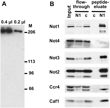
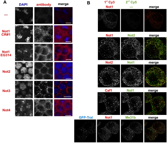
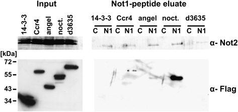
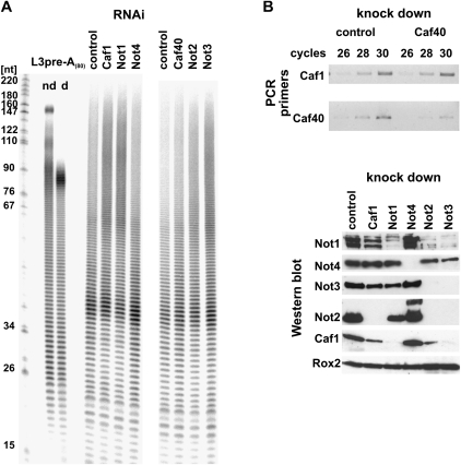
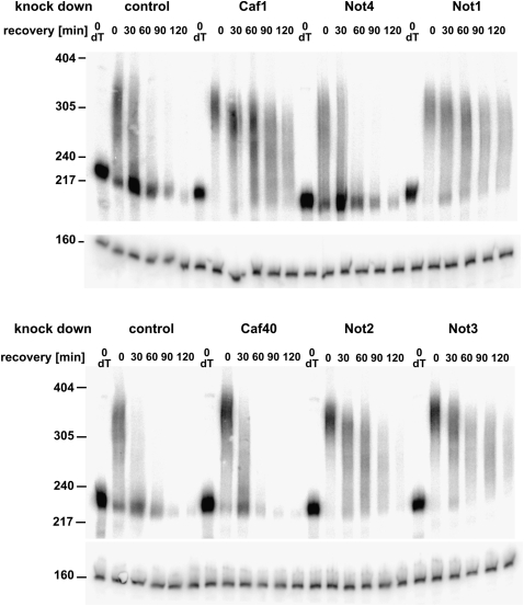
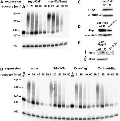
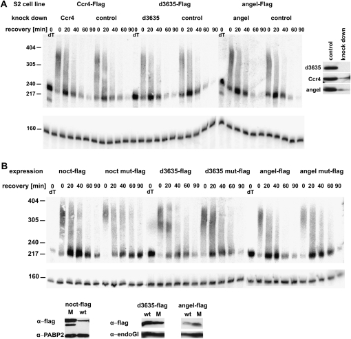
The CCR4-NOT complex is the main enzyme catalyzing the deadenylation of mRNA. We have investigated the composition of this complex in Drosophila melanogaster by immunoprecipitation with a monoclonal antibody directed against NOT1. The CCR4, CAF1 (=POP2), NOT1, NOT2, NOT3, and CAF40 subunits were associated in a stable complex, but NOT4 was not. Factors known to be involved in mRNA regulation were prominent among the other proteins coprecipitated with the CCR4-NOT complex, as analyzed by mass spectrometry. The complex was localized mostly in the cytoplasm but did not appear to be a major component of P bodies. Of the known CCR4 paralogs, Nocturnin was found associated with the subunits of the CCR4-NOT complex, whereas Angel and 3635 were not. RNAi experiments in Schneider cells showed that CAF1, NOT1, NOT2, and NOT3 are required for bulk poly(A) shortening and hsp70 mRNA deadenylation, but knock-down of CCR4, CAF40, and NOT4 did not affect these processes. Overexpression of catalytically dead CAF1 had a dominant-negative effect on mRNA decay. In contrast, overexpression of inactive CCR4 had no effect. We conclude that CAF1 is the major catalytically important subunit of the CCR4-NOT complex in Drosophila Schneider cells. Nocturnin may also be involved in mRNA deadenylation, whereas there is no evidence for a similar role of Angel and 3635.


Figures

FIGURE 1.
FIGURE 1.
Association of the CCR4-NOT polypeptides in a stable complex. (A) Detection of NOT1 with a monoclonal antibody. Indicated amounts of an extract from 0- to 2-h-old Drosophila embryos were separated on a 6% SDS–polyacrylamide gel, blotted to a PVDF membrane, and probed with the monoclonal antibody CISS 2G5. Sizes of marker proteins are indicated. (B) Western blot analysis of NOT1 immunoprecipitation from embryo extract. The monoclonal NOT1 antibody CISS 2G5 and a monoclonal rat antibody against human hnRNP K were used for immunoprecipitation (lanes labeled N1 and c, respectively). Equal amounts of input and flow-through were loaded. Bound proteins were eluted with the peptide used for immunization. A polyclonal antibody (CR#1) directed against the same peptide was used for detection of NOT1. Polyclonal antibodies for detection of the other subunits are indicated on the left. Note that the CCR4 antibody recognizes a doublet in embryo extracts. Only the lower band (arrowhead) is depleted by NOT1 immunoprecipitation and enriched in the peptide eluate. The identity of the upper band is unknown.
FIGURE 2.
FIGURE 2.
Localization of subunits of the CCR4-NOT complex. (A) Confocal images of immunostained Drosophila embryos. Blastoderm stage embryos were incubated with the antiserum indicated on the left and a Cy3-labeled secondary antibody (middle panel). DAPI was included in the mounting medium to label the nuclei (left panel). The white bar in the merge (right panel) represents 5 μm in size. Colors in the merge are blue for the DAPI staining and red for the immunostaining. (B) Colocalization of different CCR4-NOT complex subunits in S2 cells. S2 cells were sequentially stained for two different subunits of the complex. The primary antibodies used are indicated above each image. The first secondary antibody was always Cy3-labeled (left panel); the second secondary, always Cy5-labeled (middle panel). The top panel shows a control in which the second primary antibody was omitted. No Cy5 signal was seen, confirming that the first secondary antibody had been used at saturating concentrations. The bottom panel shows sequential staining for NOT1 and Me31B in cells transiently expressing GFP-Tral for labeling of P-bodies. In the merged images (right panel), the Cy3 channel is in red, the Cy5-channel in green, and GFP in blue. The overlay of red and green becomes yellow, and the overlay of green and blue becomes turquoise.
FIGURE 3.
FIGURE 3.
Developmental expression profile of the subunits of the CCR4-NOT complex. Protein extracts were from 20 embryos, 0.2 ovaries, 0.5 females, and one male. The blot was probed with the monoclonal anti-NOT1 antibody 2G5 and with polyclonal antibodies against NOT2, 3, and 4 as indicated on the left. α-Tubulin served as a loading control. The same blot was stripped and reprobed with the different antibodies.
FIGURE 4.
FIGURE 4.
Association of CCR4 and its paralogs with the CCR4-NOT complex. S2 cells were stably transformed with plasmids expressing individual members of the CCR4 family with C-terminal Flag tags under the control of a metallothionein promoter. 14-3-3 ɛ-Flag was used as a control for nonspecific association with the CCR4-NOT complex. Expression was induced for 20 h before the cells were lysed. A Western blot (left panel; 2.5% input) showed that the Flag-tagged CCR4 family members were expressed at similar levels, whereas the 14-3-3 control protein was more strongly expressed. NOT2 served as a loading control. The monoclonal NOT1 antibody CISS 2G5 was used for immunoprecipitation (lanes labeled N1), and an unrelated monoclonal antibody served as a control (lanes labeled C). Proteins were eluted with the immunogenic NOT1 peptide; 2.5% of the input and 25% of the eluate were separated on a 6% gel and a 10% SDS gel, blotted, and probed with antibodies against NOT2 (6% gel) or the Flag epitope (10% gel). Input and eluate of the Flag Western had different exposure times. The Flag-reactive band that appears to be present in the peptide eluate of the Angel expressing cells is not Angel, as the molecular weight does not match that of Angel in the left panel.
FIGURE 5.
FIGURE 5.
Involvement of individual subunits of the CCR4-NOT complex in bulk mRNA deadenylation. (A) Analysis of the length of bulk poly(A) in S2 cells after 5 d of incubation with dsRNA as indicated. DsRNA against luciferase served as a control. Total RNA was isolated from the cells, 3′-labeled, digested with RNases A and T1, and separated on a 9% polyacrylamide-urea gel. The RNA samples were from the 90-min recovery time point of Fig. 6. As a control for the specificity of the RNase digestion, in vitro synthesized L3preA(80) RNA (Kerwitz et al. 2003) was digested like the experimental RNA samples (d, digested; nd, nondigested). Lengths of DNA markers (in nucleotides) are indicated on the left. (B) Control of the knock-downs by RT-PCR (CAF40; upper panel) or Western blot (all others; lower panel). Oligo(dT)-primed reverse transcription was performed on DNase-treated RNA from knock-down and control cells. PCR primers were used to amplify either the CAF40 ORF or the CAF1 ORF as control. Aliquots of the PCR reactions were taken after 26, 28, or 30 cycles of amplification and analyzed on a 1% agarose gel. For the Western blot, control or knock-down cells were lysed in SDS sample buffer after 5 d of incubation with dsRNA and analyzed on 6% or 10% SDS polyacrylamide gels. Western blots were performed against the proteins indicated. PABP2 (Benoit et al. 1999) served as loading control.
FIGURE 6.
FIGURE 6.
Involvement of individual subunits of the CCR4-NOT complex in hsp70 mRNA deadenylation. Schneider cells treated with the double-stranded RNAs indicated were heat-shocked for 30 min at 35.5°C and then allowed to recover at 25°C. RNA was prepared at the indicated times after recovery and digested with RNase H in the presence of an hsp70-specific oligonucleotide. dT indicates Oligo(dT) was included in the RNase H digestion to mark the fully deadenylated RNA. Products were analyzed by Northern blot with probes against the 3′UTR of hsp70 and against U1 RNA serving as loading control. For control of the knock-down efficiency, see Figure 5; RNA samples were from the same cells.
FIGURE 7.
FIGURE 7.
CAF1 carries the catalytic activity for mRNA deadenylation in S2 cells. (A) Expression of myc-CAF1 or a variant containing a double point mutation in the first exonuclease motif was induced for 20 h in stably transformed S2 cells. Cells were heat-shocked for 30 min at 35.5°C and then allowed to recover at 25°C. RNA was prepared at the indicated times after recovery and digested with RNase H in the presence of an hsp70-specific oligonucleotide. dT indicates Oligo(dT) was included in the RNase H digestion to mark the fully deadenylated RNA. Products were analyzed by Northern blot with probes against the 3′UTR of hsp70 and against U1 RNA serving as loading control. (B) An equivalent experiment was carried out with cells expressing flag tagged CCR4, a CCR4 variant containing a double point mutation in the exo III domain, or a control protein. (C, D) Expression of the tagged proteins was monitored by Western blotting with the antibodies indicated. EndoGI (Temme et al. 2009) served as loading control. (E) Wild-type and mutant CCR4-Flag were precipitated with anti-Flag antibodies and eluted with Flag peptide. Equal amounts of the eluates were separated on a 10% gel, blotted, and probed with antibodies against NOT3 or CCR4.
FIGURE 8.
FIGURE 8.
Angel and 3635 do not play a detectable role in mRNA deadenylation (A) RNAi experiment: S2 cells stably transformed with expression constructs for Flag-tagged CCR4, Angel, or 3635 were treated with the corresponding dsRNA for 4 d under noninducing conditions. Then a 30-min heat shock at 35.6°C was applied. After return to 25°C, RNA was prepared at the indicated times of recovery and digested with RNase H in the presence of an hsp70-specific oligonucleotide. dT indicates Oligo(dT) was included in the RNase H digestion to mark the fully deadenylated RNA. Products were analyzed by Northern blot with probes against the 3′UTR of hsp70 and against U1 RNA serving as loading control. (Right panel) Expression of the flag-tagged proteins was induced for 4 h in small aliquots of the RNAi cells and in controls cells. Knock-down efficiency was examined by Western blot analysis with an anti-Flag antibody. Equal loading was controlled by Ponceau staining (data not shown). (B) Dominant-negative experiment: S2 cells expressing either Flag-tagged CCR4 paralogs or variants containing double point mutations in their exo III domains were grown under inducing conditions for 20 h, heat-shocked for 30 min at 35.5°C, and then allowed to recover at 25°C. RNA was prepared at the indicated times of recovery, and hsp70 mRNA was analyzed as above. RNA samples from nontransformed cells with and without addition of copper sulfate were analyzed in parallel and were indistinguishable from the samples expressing the wild-type CCR4 paralogs (data not shown). (Bottom panel) Expression of the tagged proteins was monitored by Western blotting with the antibodies indicated. EndoGI (Temme et al. 2009) and PABP2 served as loading controls.

Similar articles

Cited by

References

    1. Albert TK, Lemaire M, van Berkum NL, Gentz R, Collart M, Timmers HTM 2000. Isolation and characterization of human orthologs of yeast CCR4-NOT complex subunits. Nucleic Acids Res 28: 809–817 - PMC - PubMed
    1. Albert TK, Hanzawa H, Legtenberg YIA, de Ruwe MJ, van den Heuwel FAJ, Collart MA, Boelens R, Timmers HTM 2002. Identification of a ubiquitin-protein ligase subunit within the CCR4-NOT transcription repressor complex. EMBO J 21: 355–364 - PMC - PubMed
    1. Aslam A, Mittal S, Koch F, Andrau J-C, Winkler GS 2009. The Ccr4-Not deadenylase subunits CNOT7 and CNOT8 have overlapping roles and modulate cell proliferation. Mol Biol Cell 20: 3840–3850 - PMC - PubMed
    1. Aviv T, Lin Z, Lau S, Rendl LM, Sicheri F, Smibert CA 2003. The RNA binding SAM domain of Smaug defines a new family of post-transcriptional regulators. Nat Struct Biol 10: 614–621 - PubMed
    1. Baggs JE, Green CB 2003. Nocturnin, a deadenylase in Xenopus laevis retina: A mechanism for posttranscriptional control of circadian-related mRNA. Curr Biol 13: 189–198 - PubMed

Publication types

MeSH terms