. 2010 Aug 15;19(16):3114-23.
doi: 10.1093/hmg/ddq214. Epub 2010 May 26.

The MeCP2/YY1 interaction regulates ANT1 expression at 4q35: novel hints for Rett syndrome pathogenesis

Affiliations

The MeCP2/YY1 interaction regulates ANT1 expression at 4q35: novel hints for Rett syndrome pathogenesis

Greta Forlani et al. Hum Mol Genet. .

Abstract

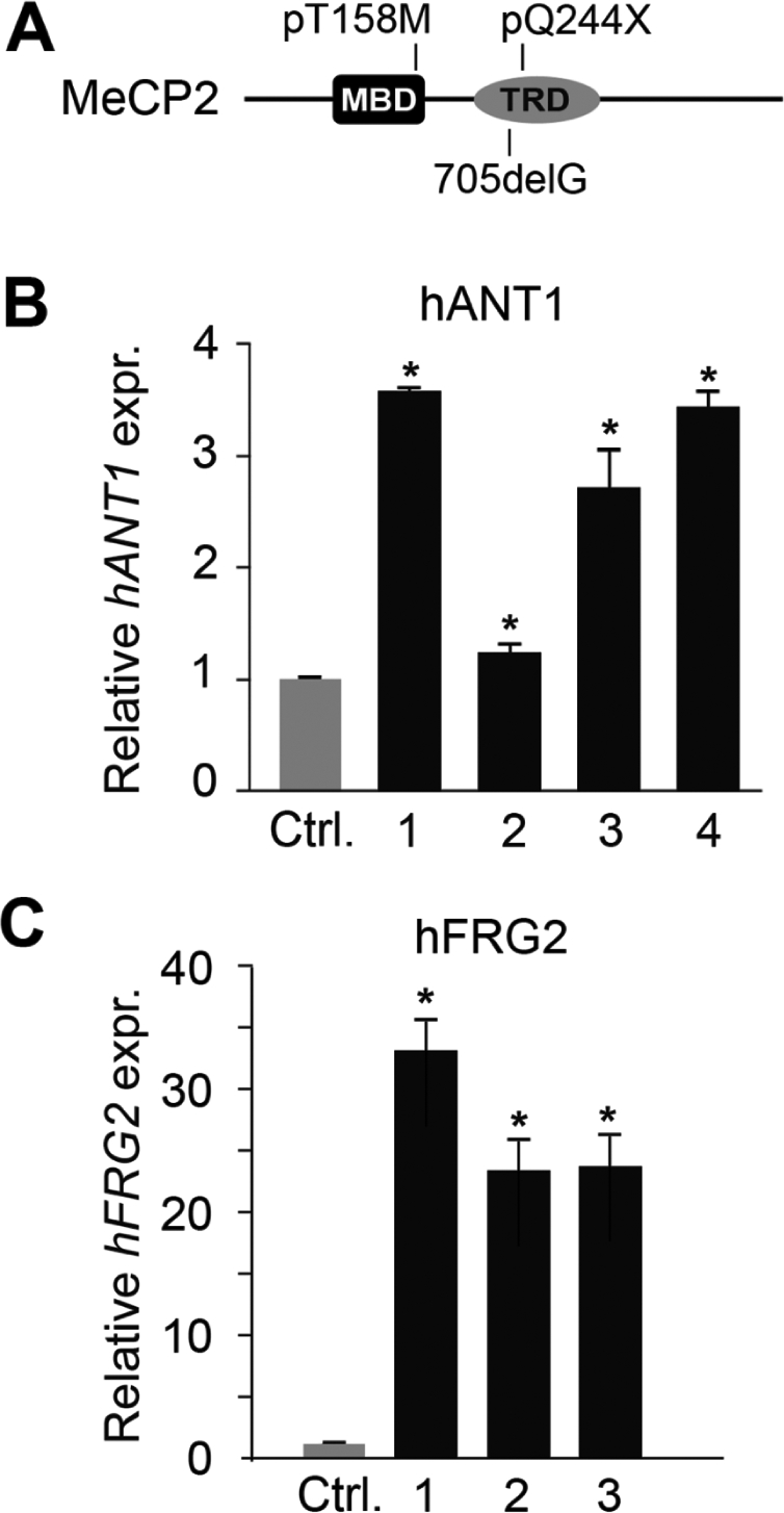
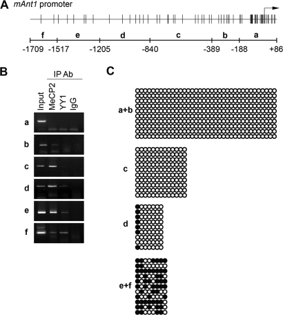
Rett syndrome is a severe neurodevelopmental disorder mainly caused by mutations in the transcriptional regulator MeCP2. Although there is no effective therapy for Rett syndrome, the recently discovered disease reversibility in mice suggests that there are therapeutic possibilities. Identification of MeCP2 targets or modifiers of the phenotype can facilitate the design of curative strategies. To identify possible novel MeCP2 interactors, we exploited a bioinformatic approach and selected Ying Yang 1 (YY1) as an interesting candidate. We demonstrate that MeCP2 interacts in vitro and in vivo with YY1, a ubiquitous zinc-finger epigenetic factor regulating the expression of several genes. We show that MeCP2 cooperates with YY1 in repressing the ANT1 gene encoding a mitochondrial adenine nucleotide translocase. Importantly, ANT1 mRNA levels are increased in human and mouse cell lines devoid of MeCP2, in Rett patient fibroblasts and in the brain of Mecp2-null mice. We further demonstrate that ANT1 protein levels are upregulated in Mecp2-null mice. Finally, the identified MeCP2-YY1 interaction, together with the well-known involvement of YY1 in the regulation of D4Z4-associated genes at 4q35, led us to discover the anomalous depression of FRG2, a subtelomeric gene of unknown function, in Rett fibroblasts. Collectively, our data indicate that mutations in MeCP2 might cause the aberrant overexpression of genes located at a specific locus, thus providing new candidates for the pathogenesis of Rett syndrome. As both ANT1 mutations and overexpression have been associated with human diseases, we consider it highly relevant to address the consequences of ANT1 deregulation in Rett syndrome.


Figures

Figure 1.
Figure 1.
MeCP2 and YY1 interact directly in vitro. (A) The MeCP2 region that binds YY1 was identified by GST pull-down assays in which full-length in vitro translated [35S]methionine-labeled MeCP2 or its mutated derivatives were incubated with immobilized recombinant GST and GST–YY1. The MeCP2 derivatives are shown schematically to the left (filled circles indicate the methionines present in MeCP2) and the corresponding autoradiograms to the right. + and − indicate the presence and absence, respectively, of interaction. (B) A far-western assay (FW) was performed with 1 µg of GST and GST–YY1, separated by SDS–PAGE, transferred to a nitrocellulose membrane and probed with recombinant MeCP2 after renaturation. MeCP2 was detected with anti-MeCP2 antibodies (lanes 1 and 2). Coomassie staining (Coom.) was used to visualize the recombinant GST and GST–YY1 proteins (lanes 3 and 4, respectively) used in the far-western assay.
Figure 2.
Figure 2.
MeCP2 and YY1 interact in vivo. (A) Co-IP of exogenously expressed Myc-MeCP2 and Flag-YY1. HEK293 cells were transiently transfected with vectors expressing Myc-MeCP2 and Flag or Flag-YY1 in different combinations. A total cell extract (TCE) was immunoprecipitated with anti-Flag antibodies, and the precipitated proteins were revealed with anti-MeCP2 (monoclonal) and anti-YY1 (polyclonal) antibodies. Inputs corresponding to 10% of the TCEs were also analyzed. (B) Co-IP of endogenous MeCP2 and YY1 from a mouse brain extract. YY1 was immunoprecipitated from a P28 mouse brain TCE and the precipitated proteins revealed with anti-MeCP2 and anti-YY1. Mouse IgGs were used as negative control. Input corresponds to 10% of the TCE. (C) Endogenous YY1 and MeCP2 partially co-localize in NIH3T3 cells. Cells were immunostained with anti-MeCP2 (a, polyclonal) and anti-YY1 (b, monoclonal) antibodies and counterstained with DAPI (d). Merged images are shown in (c).
Figure 3.
Figure 3.
ANT1 expression is upregulated in MeCP2- and YY1-depleted cells. (A) HeLa cells were transiently transfected with the indicated siRNAs; total cell extracts were collected 24 h post-transfection and analyzed by western blotting using anti-MeCP2 and anti-YY1 antibodies. (B) qRT-PCR analysis of ANT1 mRNA expression relative to Actin performed on RNA extracted from siRNA-treated HeLa cells (A). ANT1 expression in control cells transfected with a scrambled siRNA was normalized to 1 (Ctrl). Statistical analysis was performed on samples analyzed in triplicate. Significance was tested using the t-test. *P < 0.05. Error bars indicate standard deviations. This experiment is representative of three independent experiments.
Figure 4.
Figure 4.
ANT1 and FRG2 mRNAs are upregulated in RTT patients' fibroblasts. (A) Schematic illustration of the MeCP2 mutations in the analyzed Rett patients' fibroblast. (B) ANT1 mRNA levels in primary fibroblasts from four RTT patients with different MECP2 mutations were measured by qRT-PCR analysis and normalized to Actin. (C) FRG2 mRNA levels were analyzed in fibroblasts from three RTT patients by qRT-PCR analysis and normalized to B2M levels. The different bars correspond to the following MECP2 mutations: bar 1, 705delG; bar 2, pQ244X; bars 3 and 4, pT158M. ANT1 and FRG2 mRNA expression in fibroblasts from three unaffected patients (Ctrl) was normalized to 1. Statistical analysis was performed on samples analyzed in triplicate. Significance was tested using the t-test. *P < 0.05. Error bars indicate standard deviations.
Figure 5.
Figure 5.
MeCP2 and YY1 depletion increases Ant1 expression in murine cells and brain. (A) N2a cells were transiently transfected with the indicated siRNAs; total RNA was extracted 24 h post-transfection and analyzed by qRT-PCR to determine Ant1 mRNA levels relative to GAPDH. Ant1 expression in control cells transfected with a scrambled siRNA was normalized to 1 (Ctrl). Statistical analysis was performed on samples analyzed in triplicate. Significance was tested using the t-test. P < 0.05. Error bars indicate standard deviations. This experiment is representative of three independent experiments. (B) Representative qRT-PCR analysis showing Ant1 up regulation in the brain and cerebellum (Cer.) of one Mecp2-null mouse (KO). Similar results were observed with six other mutant animals in which we consistently observed a more pronounced augmention in the cerebellum. The data were evaluated using the t-test. *P < 0.05. Error bars indicate standard deviations. Ant1 mRNA levels are relative to GAPDH. Ant1 expression in the WT mouse was normalized to 1. (C) Western blot showing ANT1 protein levels in total extracts of tibial muscles from normal and Mecp2-null mice. Numbers below the western blot indicate the increase in ANT1 levels normalized to α-tubulin with respect to the wild-type controls.
Figure 6.
Figure 6.
MeCP2 and YY1 bind to a specific region of the mouse Ant1 promoter. (A) Schematic representation of the Ant1 promoter region used for PCR amplification of immunoprecipitated DNA. The sequence was amplified by PCR in six sub regions (a–f). The numbers −188, −389, −840, −1205, −1517 and −1709 indicate the relative distance upstream of the transcriptional start site (black arrow). Vertical bars in the upper part indicate CpG dinucleotides. (B) ChIP reveals binding of MeCP2 and YY1 to a common region of the Ant1 promoter. Chromatin from mouse cerebella was immunoprecipitated with anti-MeCP2 and anti-YY1 antibodies or control IgGs and analyzed by PCR. Non-precipitated chromatin was used as control (Input). (C) CpG methylation of the Ant1 promoter region. The promoter region was divided into four sub-regions (a + b, c, d and e + f) and the methylation status of CpGs monitored by bisulfite analysis. The filled and open circles represent the methylated and unmethylated CpG dinucleotides, respectively. A mean of 10 separate clones was analyzed for each region. Partial methylation was observed in d, e + f.

References

    1. Jones P.L., Veenstra G.J., Wade P.A., Vermaak D., Kass S.U., Landsberger N., Strouboulis J., Wolffe A.P. Methylated DNA and MeCP2 recruit histone deacetylase to repress transcription. Nat. Genet. 1998;19:187–191. doi:10.1038/561. - DOI - PubMed
    1. Nan X., Ng H.H., Johnson C.A., Laherty C.D., Turner B.M., Eisenman R.N., Bird A. Transcriptional repression by the methyl-CpG binding protein MeCP2 involves a histone deacetylase complex. Nature. 1998;393:386–389. - PubMed
    1. Fuks F., Hurd P.J., Deplus R., Kouzarides T. The methyl-CpG binding protein MeCP2 links DNA methylation to histone methylation. Nucleic Acids Res. 2003;31:2305–2312. doi:10.1093/nar/gkg332. - DOI - PMC - PubMed
    1. Nan X., Hou J., Maclean A., Nasir J., Lafuente M.J., Shu X., Kriaucionis S., Bird A. Interaction between chromatic proteins MECP2 and ATRX is disrupted by mutations that cause inherited mental retardation. Proc. Natl Acad. Sci. USA. 2007;104:2709–2714. doi:10.1073/pnas.0608056104. - DOI - PMC - PubMed
    1. Chahrour M., Jung S.Y., Shaw C., Zhou X., Wong S.T., Qin J., Zoghbi H.Y. MeCP2, a key contributor to neurological disease, activates and represses transcription. Science. 2008;320:1224–1229. doi:10.1126/science.1153252. - DOI - PMC - PubMed

Publication types

MeSH terms

Substances