

Changes in apparent diffusion coefficients in the normal uterus during different phases of the menstrual cycle
- PMID: 20505034
- PMCID: PMC3473588
- DOI: 10.1259/bjr/11056533
Changes in apparent diffusion coefficients in the normal uterus during different phases of the menstrual cycle
Abstract
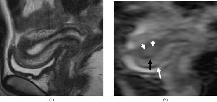
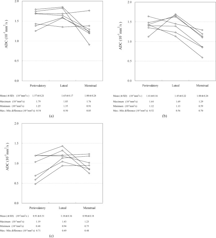
This study investigated the apparent diffusion coefficients (ADCs) of the uterine zonal structures (myometrium, endometrium and junctional zone) among reproductive women, and their changes during the menstrual cycle. Magnetic resonance (MR) images of seven healthy females (aged 24-31 years) were obtained during the periovulatory, luteal and menstrual phases. Diffusion-weighted imaging (DWI) was performed with a single-shot echo-planar imaging (EPI) sequence in the midsagittal plane of the uterus using three b-values (b = 0, 500 or 1000 s mm(-2)). The ADC values of the three uterine zonal structures were measured on an ADC map by placing two regions of interest (ROI) on the corresponding zonal structures. The average changes of ADC values (intra-individual ADC value variation) over three menstrual phases were 0.41 x 10(-3) mm(2) s(-1) (range, 0.08-0.91) for myometrium, 0.55 x 10(-3) mm(2) s(-1) (0.35-0.84) for endometrium, and 0.40 x 10(-3) mm(2) s(-1) (0.18-0.59) for the junctional zone. The ADC values for myometrium and endometrium were lower in the menstrual phase, although there was some overlap of individual values. Interindividual variation in ADC value for a given zone or phase ranged from 0.48 x 10(-3) mm(2) s(-1) to 0.85 x 10(-3) mm(2) s(-1). Intermeasurement variation between the two ROIs ranged from 0 to 0.48 x 10(-3) mm(2) s(-1) per measurement. The magnitude of these variations was comparable to reported differences between malignant and non-malignant tissues. These preliminary results, from a small number of subjects, suggest that the menstrual cycle and individual variation in pre-menopausal women should be considered when interpreting the ADC values of uterine structures.
Figures
References
-
- Le Bihan D, Turner R, MacFall JR. Effects of intravoxel incoherent motions (IVIM) in steady-state free precession (SSFP) imaging: application to molecular diffusion imaging. Magn Reson Med 1989;10:324–37 - PubMed
-
- Lyng H, Haraldseth O, Rofstad EK. Measurement of cell density and necrotic fraction in human melanoma xenografts by diffusion weighted magnetic resonance imaging. Magn Reson Med 2000;43:828–36 - PubMed
-
- Koh DM, Padhani AR. Diffusion-weighted MRI: a new functional clinical technique for tumour imaging. Br J Radiol 2006;79:633–5 - PubMed
-
- DeSouza NM, Reinsberg SA, Scurr ED, Brewster JM, Payne GS. Magnetic resonance imaging in prostate cancer: the value of apparent diffusion coefficients for identifying malignant nodules. Br J Radiol 2007;80:90–5 - PubMed
-
- Vandecaveye V, de Keyzer F, Vander Poorten V, Deraedt K, Alaerts H, Landuyt W, et al. Evaluation of the larynx for tumour recurrence by diffusion-weighted MRI after radiotherapy: initial experience in four cases. Br J Radiol 2006;79:681–7 - PubMed
