. 2010 Sep;89(9):996-1001.
doi: 10.1177/0022034510372885. Epub 2010 May 26.

Biodegradation of resin-dentin interfaces increases bacterial microleakage

Affiliations

Biodegradation of resin-dentin interfaces increases bacterial microleakage

S Kermanshahi et al. J Dent Res. 2010 Sep.

Abstract

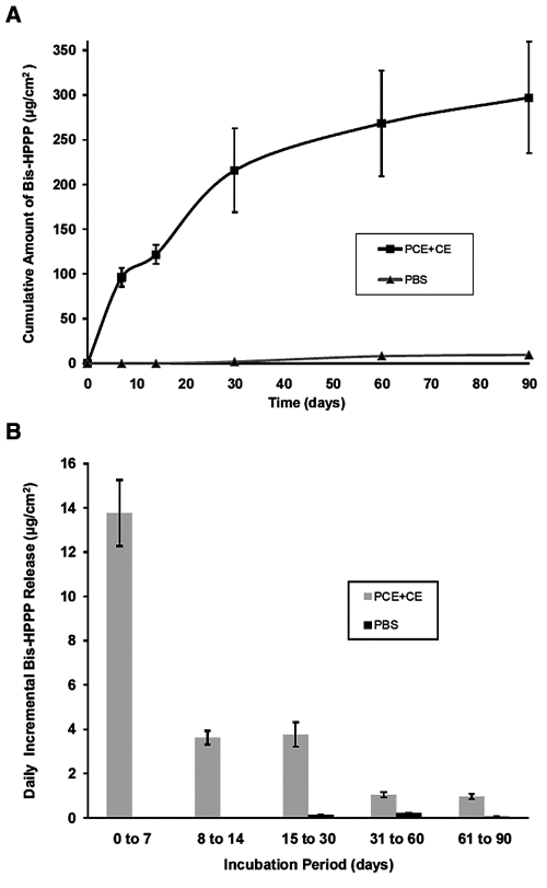
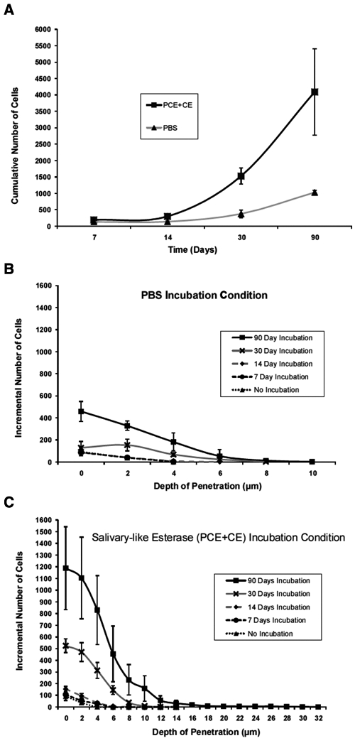
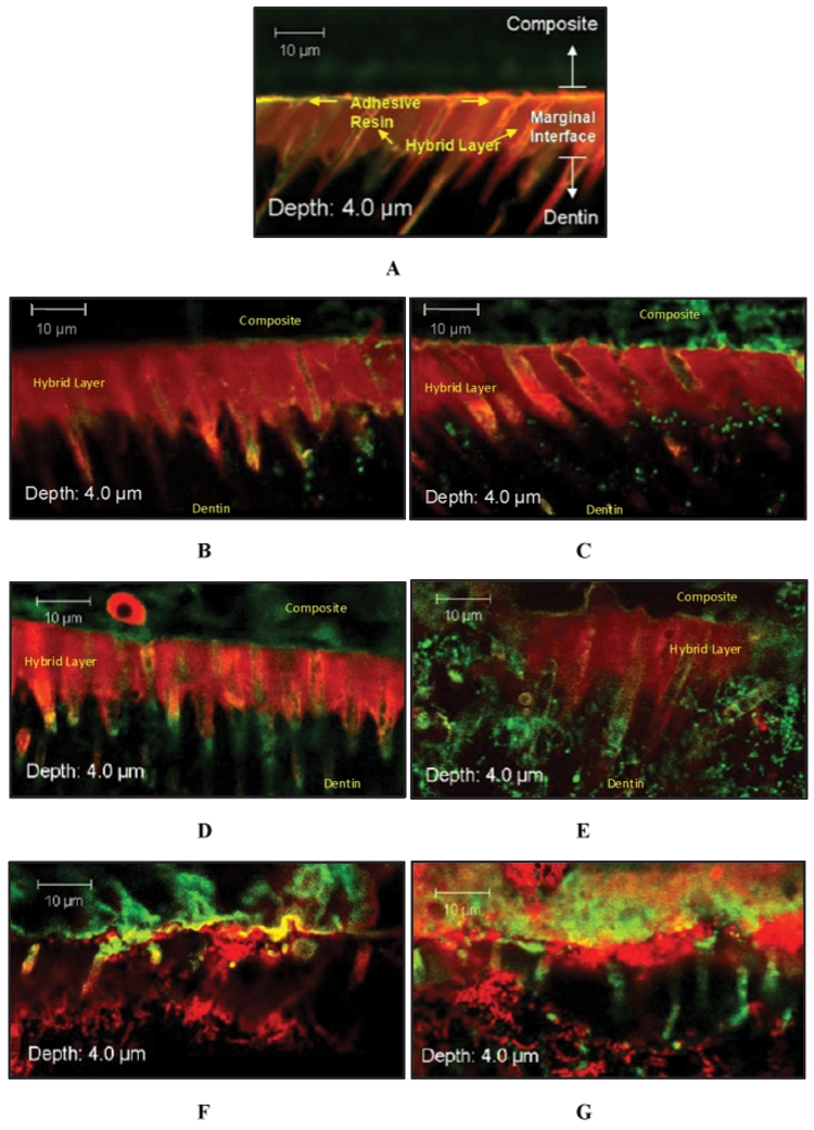
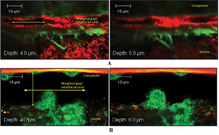
Bis-GMA-containing resin composites and adhesives undergo biodegradation by human-saliva-derived esterases, yielding Bis-hydroxy-propoxy-phenyl-propane (Bis-HPPP). The hypothesis of this study is that the exposure of dental restorations to saliva-like esterase activities accelerates marginal bacterial microleakage. Resin composites (Scotchbond, Z250, 3M) bonded to human dentin were incubated in either buffer or dual-esterase media (pseudocholinesterase/cholesterol-esterase; PCE+CE), with activity levels simulating those of human saliva, for up to 90 days. Incubation solutions were analyzed for Bis-HPPP by high-performance liquid chromatography. Post-incubation, specimens were suspended in a chemostat-based biofilm fermentor cultivating Streptococcus mutans NG8, a primary species associated with dental caries, for 7 days. Bacterial microleakage was assessed by confocal laser scanning microscopy. Bis-HPPP production and depth and spatial volume of bacterial cell penetration within the interface increased with incubation time and were higher for 30- and 90-day PCE+CE vs. buffer-incubated groups, suggesting that biodegradation can contribute to the formation of recurrent decay.


Figures

Figure 1.
Figure 1.
Bis-HPPP release from resin-dentin specimens. (A) Cumulative amount of Bis-HPPP produced from resin-dentin specimens incubated in PCE+CE or PBS buffer for 7, 14, 30, and 90 days (pH 7, 37°C). (B) Incremental amount of Bis-HPPP produced from resin-dentin specimens incubated in PCE+CE or PBS buffer. All data are reported with standard error of the mean (n = 3).
Figure 2.
Figure 2.
Bacterial penetration along the resin-dentin marginal interface. (A) Cumulative numbers of bacterial cells found penetrating the marginal interface for PBS controls and PCE+CE-incubated specimens over time. Number of cells vs. depth of penetration at interfacial ROIs of (B) PBS-incubated and (C) PCE+CE-incubated specimens for 0, 7, 14, 30, and 90 days (pH 7, 3°C). All data are reported with standard error of the mean (n = 3).
Figure 3.
Figure 3.
Selected Z-stack image series captured from interfacial margins of resin-dentin specimens assigned to either (A) non-incubated, (B) 7-day PBS incubation, (C) 7-day PCE+CE incubation, (D) 30-day PBS incubation, (E) 30-day PCE+CE incubation, (F) 90-day PBS incubation, or (G) 90-day PCE+CE incubation. Interfacial zones (composite, adhesive, hybrid layer, and dentin) are distinguishable in A and, to a lesser extent, in B-E; however, in F and G, the organization of these marginal components is disrupted. Resin impregnation of dentinal tubules in the hybrid layer is disrupted (F and G). Specimens were stained by means of a Live/Dead Baclight Viability Kit (magnification X62, 2X zoom). Live cells indicated by green fluorescence through interaction with Syto9; dead cells indicated by red fluorescence through interaction with propidium iodide.
Figure 4.
Figure 4.
Selected Z-stack image series captured at interfacial ROIs of 2 90-day PCE+CE-incubated resin-dentin specimens. (A) Interfacial void spanning approximately 4-5 µm in height. (B) Interfacial void spanning over 20 µm in height. Characteristic of three-dimensional biofilm growth are interstitial voids that can be seen among fluorescently stained S. mutans microcolonies. In (B), large mushroom-shaped biofilm structures are found colonizing both the top and bottom axial walls. Specimens were stained by means of a Live/Dead Baclight Viability Kit (magnification X62, 2X zoom). Live cells indicated by green fluorescence through interaction with Syto9; dead cells indicated by red fluorescence through interaction with propidium iodide.

References

    1. Bouillaguet S. (2004). Biological risks of resin-based materials to the dentin-pulp complex. Crit Rev Oral Biol Med 15:47-60 - PubMed
    1. Cvitkovitch DG, Li YH, Ellen RO. (2003). Quorum sensing and biofilm formation in Streptococcal infections. J Clin Invest 112:1626-1632 - PMC - PubMed
    1. Donmez N, Belli S, Pashley DH, Tay FR. (2005). Ultrastructural correlates of in vivo/in vitro bond degradation in self-etch adhesives. J Dent Res 84:355-359 - PubMed
    1. Ferracane JL. (2006). Hygroscopic and hydrolytic effects in dental polymer networks. Dent Mater 22:221-222 - PubMed
    1. Finer Y, Santerre JP. (2003). Biodegradation of a dental composite by esterases: dependence on enzyme concentration and specificity. J Biomater Sci Polym Ed 14:837-849 - PubMed

Publication types

MeSH terms

LinkOut - more resources