. 2010 Jul 15;21(14):2483-99.
doi: 10.1091/mbc.e10-03-0176. Epub 2010 May 26.

Scyl1 facilitates nuclear tRNA export in mammalian cells by acting at the nuclear pore complex

Affiliations

Scyl1 facilitates nuclear tRNA export in mammalian cells by acting at the nuclear pore complex

Shawn C Chafe et al. Mol Biol Cell. .

Abstract

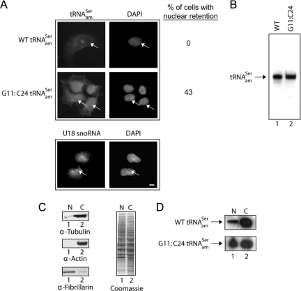
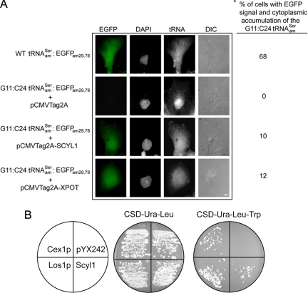
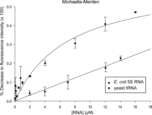
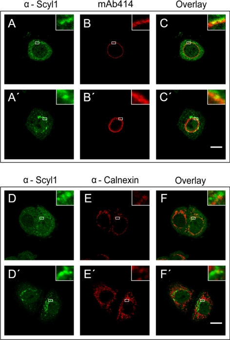
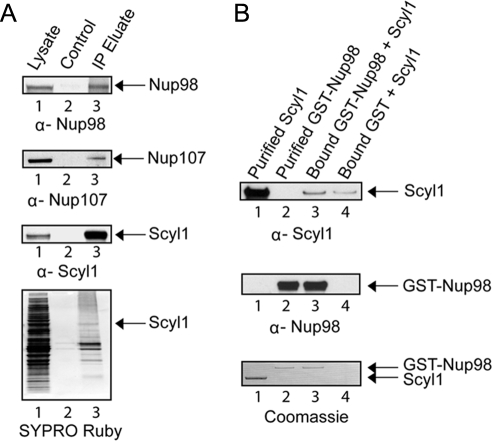
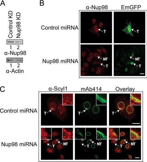
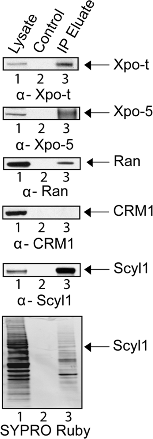
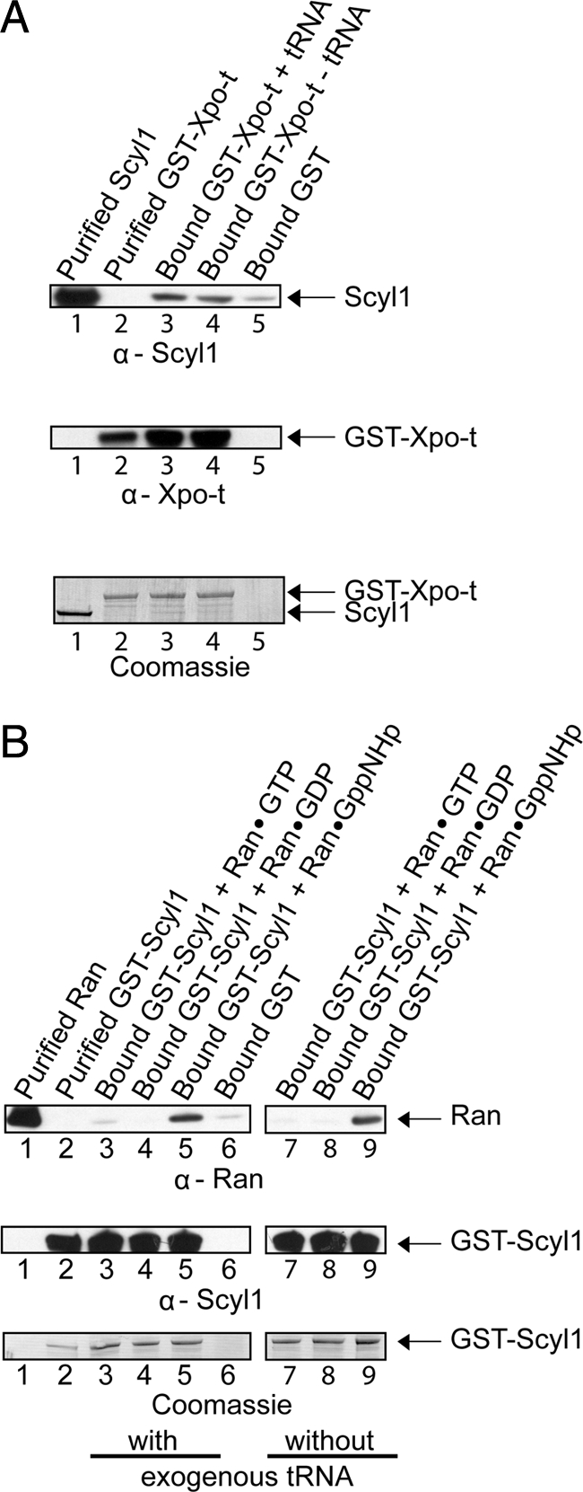
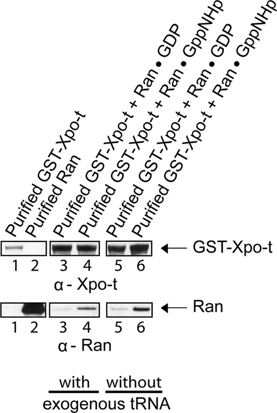
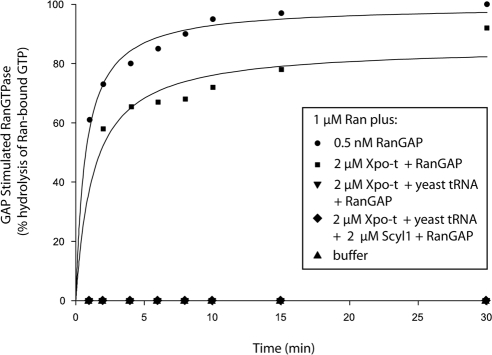
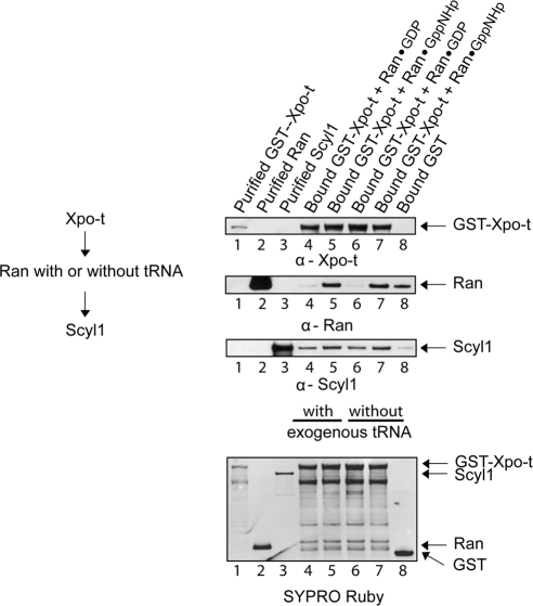
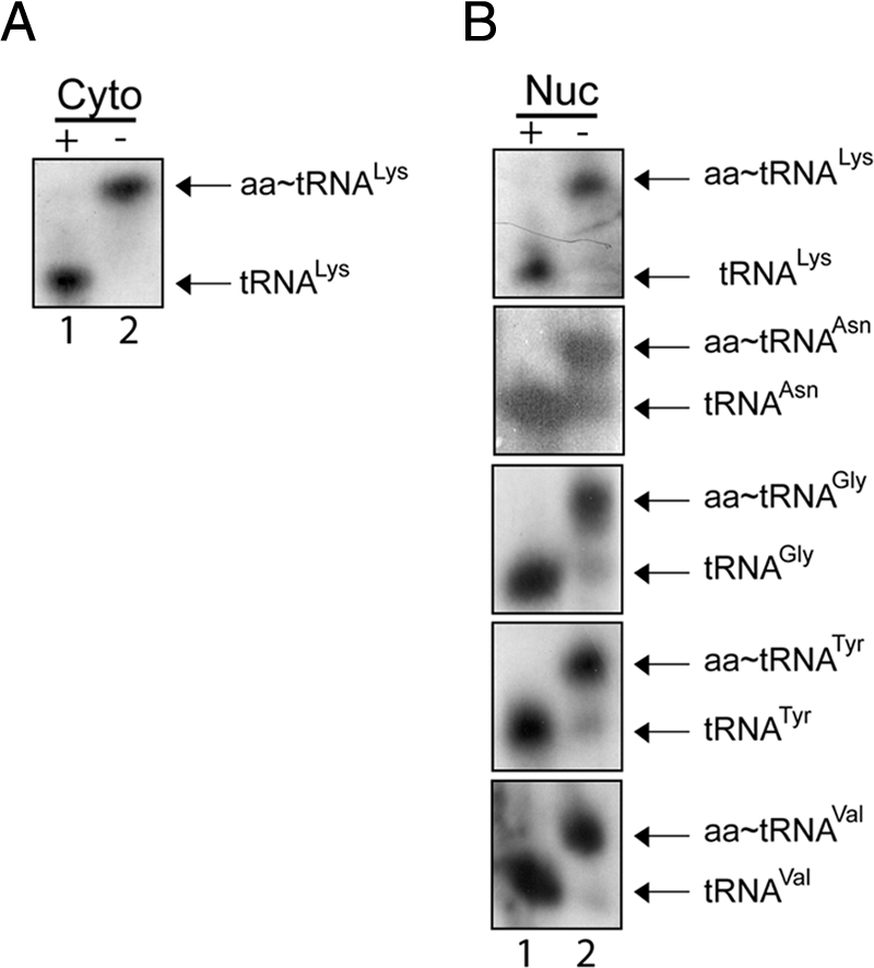
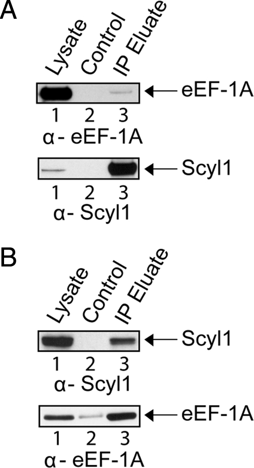
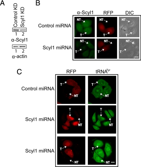
Scyl1 is an evolutionarily conserved N-terminal protein kinase-like domain protein that plays a role in COP1-mediated retrograde protein trafficking in mammalian cells. Furthermore, loss of Scyl1 function has been shown to result in neurodegenerative disorders in mice. Here, we report that Scyl1 is also a cytoplasmic component of the mammalian nuclear tRNA export machinery. Like exportin-t, overexpression of Scyl1 restored export of a nuclear export-defective serine amber suppressor tRNA mutant in COS-7 cells. Scyl1 binds tRNA saturably, and associates with the nuclear pore complex by interacting, in part, with Nup98. Scyl1 copurifies with the nuclear tRNA export receptors exportin-t and exportin-5, the RanGTPase, and the eukaryotic elongation factor eEF-1A, which transports aminoacyl-tRNAs to the ribosomes. Scyl1 interacts directly with exportin-t and RanGTP but not with eEF-1A or RanGDP in vitro. Moreover, exportin-t containing tRNA, Scyl1, and RanGTP form a quaternary complex in vitro. Biochemical characterization also suggests that the nuclear aminoacylation-dependent pathway is primarily responsible for tRNA export in mammalian cells. These findings together suggest that Scyl1 participates in the nuclear aminoacylation-dependent tRNA export pathway and may unload aminoacyl-tRNAs from the nuclear tRNA export receptor at the cytoplasmic side of the nuclear pore complex and channels them to eEF-1A.


Figures

Figure 1.
Figure 1.
The G11:C24 mutation affects the efficiency of nuclear export of tRNAamSer. (A) FISH analysis of the nuclear-cytoplasmic distribution of the WT and G11:C24 mutant amber suppressor tRNA. COS-7 cells were transfected with pSVBpUC containing the WT tRNAamSer or G11:C24 tRNAamSer gene. The subcellular distribution of the wild-type tRNAamSer and G11:C24 tRNAamSer mutant and the location of U18 snoRNA were detected using Cy3- and fluorescein-labeled oligonucleotides, respectively. The values presented are averages of three independent experiments. DNA was visualized by staining with DAPI. Bar, 10 μm. (B) The G11:C24 mutation did not affect maturation of tRNAamSer. Total RNA was isolated from COS-7 cells transfected with pSVBpUC carrying the WT (lane 1) or G11:C24 mutant tRNAamSer gene (lane 2). An aliquot containing 20 μg of total RNA was subjected to PAGE under denaturing conditions, and tRNAamSer was detected by Northern blot analysis. (C) Analyses of the purity of nuclei isolated from COS-7 cells. Nuclear (N) and cytoplasmic (C) fractions were prepared from COS-7 cells, and Western blot analysis was used to monitor the amount of the cytoplasmic markers tubulin (left, first row) and actin (second row) associated with nuclei, and the amount of the nucleolar marker fibrillarin present in the cytoplasmic fraction (third row). The amount of protein in the two fractions was assessed by Coomassie Blue staining of the blot (right). (D) Northern blot analysis of the nuclear-cytoplasmic distribution of the WT and G11:C24 mutant tRNAamSer. N and C fractions were isolated from COS-7 cells transfected with pSVBpUC carrying the G11:C24 mutant or WT suppressor tRNA gene. Total RNA as isolated from each fraction and subjected to PAGE under denaturing conditions. The RNA was transferred electrophoretically to membrane, and Northern analysis was performed to detect tRNAamSer.
Figure 2.
Figure 2.
Overexpression of Scyl1 rescues export of the yeast and human mutant amber suppressor tRNAs defective in nuclear export. (A) Scyl1 increased the efficiency of nuclear export of the export-defective human serine amber suppressor tRNASer mutant. COS-7 cells grown on coverslips were transfected with pSVBpUC-tRNAamSer-EGFPam29,78, pSVBpUC-G11:C24 tRNAamSer-EGFPam29,78, and pCMVTag2A, pSVBpUC-G11:C24 tRNAamSer-EGFPam29,78, and pCMV-XPOT, or pSVBpUC-G11:C24 tRNAamSer-EGFPam29,78 and pCMV-SCYL1, and allowed to express for 24 h. The subcellular distribution of the WT tRNAamSer and the G11:C24 tRNAamSer was detected by FISH using Cy3-labeled oligonucleotides. Expression of EGFP was monitored by fluorescent microscopy. The DNA was visualized by DAPI staining. The asterisk (*) denotes the percentage of cells expressing EGFP, and the values presented are averages of three independent experiments. For each experiment, 100 cells were analyzed for EGFP expression. Bar, 10 μm. (B) Scyl1 increased the efficiency of nuclear export of the export-defective S. cerevisiae tyrosine amber suppressor tRNA mutant. The HEY301-129 strain containing the yEPLAC195-G11:C24 tRNAamTyr plasmid was transformed with pYX242-LOS1, pYX242-CEX1, pYX242-SCYL1, or pYX242 and streaked on CSD-Ura-Leu or CSD-Ura-Leu-Trp to select for amber suppression of the trp1 allele.
Figure 3.
Figure 3.
Scyl1 binds tRNA directly and saturably. Substrate induced intrinsic fluorescence quenching of tryptophan residues was used to determine whether purified Scyl1 binds tRNA (•) or E. coli 5S RNA (■) directly in vitro as described in Materials and Methods.
Figure 4.
Figure 4.
Scyl1 associates specifically with the NPC in HeLa cells. HeLa cells were fixed with 4% paraformaldehyde in PBS and then permeabilized with 0.5% Triton X-100 in PBS (first and third rows) or fixed and permeabilized simultaneously in 2% paraformaldehyde and 0.5% Triton X-100 (second and fourth rows). The location of Scyl1, the NPC and Calnexin was detected by immunofluorescence microscopy. Localization of Scyl1 with the NPC or calnexin was determined by overlay analyses of the micrographs. Insets represent zoomed in regions outlined with white rectangles. Bars, 10 μm.
Figure 5.
Figure 5.
Scyl1 associates with the NPC by interacting directly with Nup98. (A) Nup98 copurifies with Scyl1. Total cell lysates prepared from HeLa cells (lane 1) were incubated with Sepharose 4B (lane 2) or α-Scyl1 coupled to Sepharose 4B (lane 3). The washed resins were boiled in sample buffer, and the eluates were subjected to Western blot analysis to detect Nup98 (first row), Nup107 (second row), and Scyl1 (third row). The proteins were visualized directly by SYPRO Ruby staining of the blot (fourth row). (B) Scyl1 interacts directly with Nup98 in vitro. Bound GST-Nup98 (20 μg; 161 pmol) (lane 3) or GST (lane 4) was incubated with a twofold molar excess of Scyl1 (28 μg; 322 pmol). The washed resins were boiled in sample buffer, and the eluates and purified Scyl1 (lane 1) and GST-Nup98 (lane 2) were subjected to SDS-PAGE. Scyl1 (first row) and GST-Nup98 (second row) were detected by Western blot analysis followed by Coomassie Blue staining of the blot (third row).
Figure 6.
Figure 6.
Knockdown of Nup98 expression affects the localization of Scyl1 to the NPC in HeLa cells. (A) Nup98 level is reduced upon expression of Nup98 miRNA. HeLa cells were transfected with either the control (lane 1) or Nup98 miRNA (lane 2) expression plasmid. Cell lysates were prepared 72 h after transfection, and Western blot analysis was performed to monitor Nup98 (top row) or actin (bottom row) level. (B) Reduction in the level of Nup98 is detected in vivo by fluorescence microscopy. HeLa cells were transfected with either the control (top) or Nup98 (bottom) miRNA expression plasmid. After 72 h, the cells were fixed with 4% paraformaldehyde and the level of Nup98 (left column) was monitored by immunofluorescence microscopy. Transfected cells were identified by the expression of EmGFP (right column). T, transfected cells; NT, untransfected cells. The transfection efficiency was determined by analyzing 100 cells from three independent transfections. Twenty-five transfected cells in two independent transfections were analyzed for the level of Nup98 by measuring the fluorescence intensity using the MetaMorph software. (C) A decrease in the level of Nup98 affects the localization of Scyl1 to the NPC. HeLa cells were transfected with either the control (top) or Nup98 (bottom) miRNA expression plasmid. After 72 h, the cells were fixed with 2% paraformaldehyde and permeabilized simultaneously with 0.5% Triton X-100; localization of Scyl1 (left column) was monitored by immunofluorescence microscopy. The NPC was visualized by staining with mAb414 (middle). Overlay analysis was performed to monitor the level of Scyl1 at the NPC (right) in 25 transfected cells (left box and inset) and untransfected cells (right box and inset). Insets represent zoomed in regions outlined with white rectangles. T, transfected cells; NT, untransfected cells. Bars, 10 μm.
Figure 7.
Figure 7.
Xpo-t, Xpo-5, and Ran but not Crm1 copurify with Scyl1. Total cell lysate from HeLa cells (lane 1) was incubated with Sepharose 4B (lane 2) or α-Scyl1 coupled to Sepharose 4B (lane 3). The resins were washed and boiled in sample buffer to release bound proteins. Western blot analysis was used to detect Xpo-t (first row), Xpo-5 (second row), Ran (third row), Crm1 (fourth row), and Scyl1 (fifth row) in the eluates. The proteins were visualized directly by SYPRO Ruby staining of the blot (sixth row).
Figure 8.
Figure 8.
Scyl1 interacts directly with Xpo-t and RanGTP but not RanGDP in vitro. (A) Scyl1 interacts with Xpo-t in a tRNA-independent manner. GST bound to GT-Sepharose (lane 5) or GST-Xpo-t (20 μg; 158 pmol) bound to GT-Sepharose with (lane 3) or without (lane 4) 6 μM yeast mature tRNA was incubated with a twofold molar excess of Scyl1 (27 μg; 316 pmol). The washed resins were boiled in sample buffer, and the eluates and purified Scyl1 (lane 1) and GST-Xpo-t (lane 2) were separated by SDS-PAGE. Scyl1 (first row) and GST-Xpo-t (second row) were detected by Western blot analysis followed by Coomassie Blue staining of the blot (third row). (B) Scyl1 interacts with RanGTP but not RanGDP. GST-Scyl1 (20 μg; 178 pmol) bound to GT-Sepharose in the presence (lanes 3–5) or absence (lanes 7–9) of 70 μM yeast mature tRNA and with a twofold molar excess of Ran (9 μg, 356 pmol) loaded with 100 μM GTP (lanes 3 and 7), GDP (lanes 4 and 8), or GppNHp (lanes 5 and 9). The same amount of Ran was incubated with bound GST (lane 6). The resins were washed and boiled in sample buffer to elute bound proteins. Ran (lane 1), GST-Scyl1 (lane 2), and the eluates were separated by SDS-PAGE, followed by Western blot analyses to detect Ran (first row) and GST-Scyl1 (second row). Coomassie Blue staining of the blot was used to detect the protein directly (third row).
Figure 9.
Figure 9.
Xpo-t interacts directly with RanGTP but not RanGDP in a tRNA-independent manner. GST-Xpo-t (20 μg; 158 pmol) bound to GT-Sepharose in the presence (lanes 3 and 4) or absence (lanes 5 and 6) of an excess of yeast mature tRNA (6 μM) was incubated with Ran (8 μg; 316 pmol) in the GDP (lanes 3 and 5) or RanGppNHp (lanes 4 and 6) form. The resins were washed and boiled in sample buffer to elute bound proteins. GST-Xpo-t (lane 1), Ran (lane 2), and the eluates were subjected to SDS-PAGE, followed by Western blot analyses to detect GST-Xpo-t (top row) and Ran (bottom row).
Figure 10.
Figure 10.
Xpo-t blockage of RanGAP stimulation of the GTPase activity of Ran is dependent on the presence of tRNA and is not alleviated by the inclusion of Scyl1. Ran-[γ-32P]GTP (1 μM) was incubated with (•) or without (▴) 0.5 nM RanGAP. Ran also was incubated with RanGAP in the presence of 2 μM Xpo-t with (▾) or without (■) 6 μM yeast mature tRNA, or with 2 μM Xpo-t, 2 μM Scyl1, and 6 μM yeast mature tRNA (♦). At the specified times, hydrolysis of Ran-bound GTP was determined by counting released [32P]phosphate by using the charcoal method.
Figure 11.
Figure 11.
Scyl1, Xpo-t containing endogenous E. coli tRNA and RanGTP form a complex. GST-Xpo-t (20 μg; 158 pmol) bound to GT-Sepharose was incubated in the presence (lanes 4 and 5) or absence (lanes 6 and 7) of an excess of exogenous yeast mature tRNA (6 μM) and Ran (4 μg; 158 pmol) in the GDP (lanes 4 and 6) or GppNHp (lanes 5 and 7) bound form. The resins were washed and incubated with Scyl1 (14 μg; 158 pmol). The same amount of Ran and Scyl1 was incubated with bound GST (lane 8). The resins were washed and boiled in sample buffer to elute bound proteins. GST-Xpo-t (lane 1), Ran (lane 2), Scyl1 (lane 3) and the eluates were subjected to SDS-PAGE, followed by Western blot analyses to detect GST-Xpo-t (first row), Ran (second row), and Scyl1 (third row). The proteins also were detected directly by SYPRO Ruby staining of the blot (fourth row).
Figure 12.
Figure 12.
Aminoacylated tRNAs are present in the nucleus of HeLa cells. Total RNA was isolated from nuclear and cytosolic fractions prepared from HeLa cells under acidic conditions. RNA treated with (+) or without (−) base was separated on a 6.5% acid-urea polyacrylamide gel. Northern blot analysis was performed to monitor the aminoacylation status of tRNALys in the cytoplasmic (A) and various tRNA isoacceptors in the nuclear (B) fractions using 32P-labeled oligonucleotide probes.
Figure 13.
Figure 13.
eEF-1A and Scyl1 copurify with each other. (A) eEF-1A copurifies with Scyl1. HeLa cell lysates (lane 1) were incubated with Sepharose (lane 2) or α-Scyl1-Sepharose (lane 3). The resins were washed and boiled in sample buffer to elute bound proteins. Western blot analysis was used to detect eEF-1A (first row) and Scyl1 (second row). (B) Scyl1 copurifies with eEF-1A. HeLa cell lysates (lane 1) were incubated with Sepharose (lane 2) or α-eEF-1A-Sepharose (lane 3). The resins were washed and boiled in sample buffer to elute bound proteins. Western blot analysis was used to detect Scyl1 (first row) and eEF-1A (second row).
Figure 14.
Figure 14.
Knockdown of Scyl1 expression does not affect nuclear tRNA export. (A) Scyl1 levels are reduced upon expression of a Scyl1 miRNA. HeLa cells were transfected with either the control or Scyl1 miRNA expression plasmid. Cell lysates were prepared 72 h after transfection and Western blot analysis was performed to monitor Scyl1 (top row) and actin (bottom) levels. (B) A reduction in Scyl1 levels can be detected in vivo. HeLa cells were transfected with either a control (top) or Scyl1 (bottom) miRNA expression plasmid. After 72 h, the cells were fixed and the levels of Scyl1 (left column) were monitored by immunofluorescence microscopy. Transfected cells were identified by expression of RFP (middle column), and the efficiency of transfection was determined by analyzing 100 cells from three independent transfections. Twenty-five transfected cells in two independent transfections were analyzed for the level of Scyl1 by measuring the fluorescence intensity by using the MetaMorph software. (C) Decreased Scyl1 expression did not affect the nuclear-cytoplasmic distribution of tRNA. HeLa cells were transfected with either a control or Scyl1 miRNA expression plasmid. The cellular location of tRNATyr in 25 transfected cells was detected using an oligonucleotide probe conjugated to fluorescein. Cells expressing the control (first row) and Scyl1 (second and third rows) miRNAs were visualized by confocal laser scanning microscopy. T, transfected cells; NT, untransfected cells. Bars, 10 μm.

Similar articles

Cited by

References

    1. Arts G. J., Fornerod M., Mattaj I. W. Identification of a nuclear export receptor for tRNA. Curr. Biol. 1998a;8:305–314. - PubMed
    1. Arts G. J., Kuersten S., Romby P., Ehresmann B., Mattaj I. W. The role of exportin-t in selective nuclear export of mature tRNAs. EMBO J. 1998b;17:7430–7441. - PMC - PubMed
    1. Azad A. K., Stanford D. R., Sarkar S., Hopper A. K. Role of nuclear pools of aminoacyl-tRNA synthetases in tRNA nuclear export. Mol. Biol. Cell. 2001;12:1381–1392. - PMC - PubMed
    1. Belgareh N., et al. An evolutionarily conserved NPC subcomplex, which redistributes in part to kinetochores in mammalian cells. J. Cell Biol. 2001;154:1147–1160. - PMC - PubMed
    1. Bernstein K. A., Baserga S. J. The small subunit processome is required for cell cycle progression at G1. Mol. Biol. Cell. 2004;15:5038–5046. - PMC - PubMed

Publication types

MeSH terms

LinkOut - more resources