. 2010 Jul 15;21(14):2384-98.
doi: 10.1091/mbc.e09-07-0637. Epub 2010 May 26.

Unrestrained spindle elongation during recovery from spindle checkpoint activation in cdc15-2 cells results in mis-segregation of chromosomes

Affiliations

Unrestrained spindle elongation during recovery from spindle checkpoint activation in cdc15-2 cells results in mis-segregation of chromosomes

Chuan Chung Chai et al. Mol Biol Cell. .

Abstract

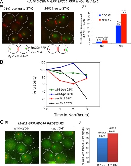
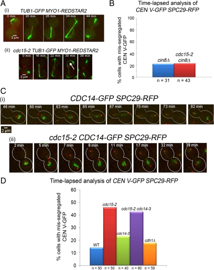
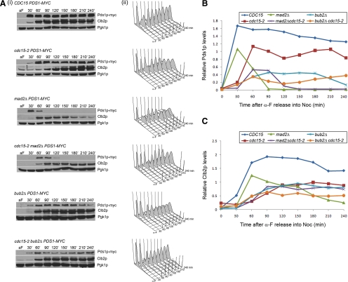
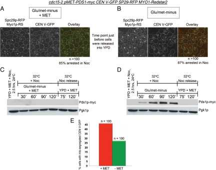
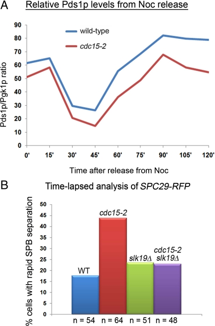
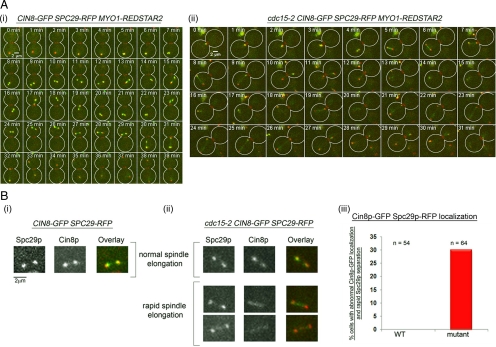
During normal metaphase in Saccharomyces cerevisiae, chromosomes are captured at the kinetochores by microtubules emanating from the spindle pole bodies at opposite poles of the dividing cell. The balance of forces between the cohesins holding the replicated chromosomes together and the pulling force from the microtubules at the kinetochores result in the biorientation of the sister chromatids before chromosome segregation. The absence of kinetochore-microtubule interactions or loss of cohesion between the sister chromatids triggers the spindle checkpoint which arrests cells in metaphase. We report here that an MEN mutant, cdc15-2, though competent in activating the spindle assembly checkpoint when exposed to Noc, mis-segregated chromosomes during recovery from spindle checkpoint activation. cdc15-2 cells arrested in Noc, although their Pds1p levels did not accumulate as well as in wild-type cells. Genetic analysis indicated that Pds1p levels are lower in a mad2Delta cdc15-2 and bub2Delta cdc15-2 double mutants compared with the single mutants. Chromosome mis-segregation in the mutant was due to premature spindle elongation in the presence of unattached chromosomes, likely through loss of proper control on spindle midzone protein Slk19p and kinesin protein, Cin8p. Our data indicate that a slower rate of transition through the cell division cycle can result in an inadequate level of Pds1p accumulation that can compromise recovery from spindle assembly checkpoint activation.


Figures

Figure 1.
Figure 1.
cdc15-2 cells exhibit CEN V-GFP segregation defects when exposed to nocodazole (Noc). (Ai) Logarithmic cultures of two CEN V-GFP SPC29-RFP MYO1-Redstar2 and cdc15-2 CEN V-GFP SPC29-RFP MYO1-Redstar2 cells at 24°C were shifted directly to 37°C for 5 h (left) or exposed to Noc (15 μg/ml) for 5 h before releasing into 37°C for 60 min. (Aii) Bar chart representing the percentage of cells with mis-segregated CEN V-GFP spots. (B) Logarithmic cultures of wild-type and cdc15-2 cells were diluted to OD600 = 0.1 and exposed to 15 μg/ml Noc for varying lengths of time. Viability was taken as a percentage of the number of colonies formed after exposure to Noc to the number of colonies formed in the untreated control (0 h). (C) MAD2-GFP NDC80-Redstar2 and cdc15-2 MAD2-GFP NDC80-Redstar2 cells were examined at the Noc-arrested stage (i) for the percentage of cells showing Mad2p-GFP signals (ii).
Figure 2.
Figure 2.
cdc15-2 cells show lagging Ndc80p signals during chromosome segregation after release from Noc. (A) cdc20Δ GAL-CDC20 NDC80-GFP SPC29-RFP (i) and cdc15-2 cdc20Δ GAL-CDC20 NDC80-GFP SPC29-RFP (ii) cells were arrested in YPD for 2.5 h at 24°C and YPD for 2.5 h at 32°C before being transferred into SC/Raff/Gal and imaged on the heated stage. The percentages of cells showing abnormal Ndc80p-GFP segregation are shown in panel iii. (B) The CDC15 (i) and cdc15-2 (ii) strains were treated similarly as in A except that Noc was added during the arrest stage in YPD. The percentages of cells showing abnormal Ndc80p-GFP segregation are shown in panel iii.
Figure 3.
Figure 3.
Altered dynamics of Spc29p-RFP separation in cdc15-2 cells released from Noc. (A) Wild-type tetR-GFP CEN5∷tetO2X112 SPC29-RFP MYO1-Redstar2 (i) and cdc15-2 tetR-GFP CEN5∷tetO2X112 SPC29-RFP MYO1-Redstar2 (ii) cells were arrested in Noc and released into 32°C, and time-lapse imaging carried out to examine the cells. Counts of cells with mis-segregated CEN V-GFP spots represented as a percentage of total cells examined in time-lapse microscopy are shown in panel iii. (B) The dynamics of Spc29p-RFP separation in five typical wild-type (Ai) and five typical cdc15-2 (Aii) cells were determined by measuring the distance between Spc29p-RFP and plotted over time.
Figure 4.
Figure 4.
Restraining spindle elongation in cdc15-2 cdc20Δ GAL-CDC20 tetR-GFP CEN5∷ tetO2X112 SPC29-RFP resulted in proper biorientation of CEN V-GFP. Wild-type (i) and cdc15-2 (ii) cells were arrested in YPD + Noc and released into SC/Glu to maintain cells in metaphase as they recover from Noc during time-lapse imaging. (iii) Percentage of cells showing proper biorientation. (iv) Plots show the time taken to biorientate CEN V-GFP in CDC15 and cdc15-2 cells. Time needed for biorientation was taken as the time between Spc29p-RPF separation, and CEN V-GFP stretched across the separated Spc29p-RFP.
Figure 5.
Figure 5.
(A) Abnormal microtubule dynamics in cdc15-2 cells released from Noc. Wild-type (i) and mutant (ii) cells carrying TUB1-GFP MYO1-REDSTAR2 were arrested in Noc, released into 32°C, and examined using time-lapse microscopy. Selected time points are shown. (B) CIN8 deletion rescued CEN V-GFP mis-segregation in cdc15-2 cells. cin8Δ tetR-GFP CEN5∷tetO2X112 SPC29-RFP and cdc15-2 cin8Δ tetR-GFP CEN5∷tetO2X112 SPC29-RFP cells were treated as described in Figure 4 and the number of cells with mis-segregated CEN V-GFP are represented as a percentage of the total number of cells counted. (C) Cdc14p-GFP is not prematurely released in cdc15-2 cells. CDC15 CDC14-GFP SPC29-RFP (i) and cdc15-2 CDC14-GFP SPC29-RFP (ii) cells were treated as described in Figure 4. (D) Mis-segregation of CEN V-GFP in cdc15-2 cells not rescued by cdc14-3. Strains with various genotypes as indicated were treated as described in Figure 3, and the number of cells with mis-segregated CEN V-GFP are represented as a percentage of the total number of cells counted.
Figure 6.
Figure 6.
Pds1p levels do not accumulate as high in cdc15-2 cells. (A) Cells from various strains were sampled at 30 min-intervals after release from α-factor into Noc at 32°C for 4 h. Western blot analysis of Pds1p-myc, Clb2p, and loading control Pgk1p are shown in panel i. FACs profiles of the cells at each time point are shown in panel ii. (B) Densitometry readings of the Western blot showing the ratio of Pds1p levels to Pgk1p levels. (C) Densitometry readings of the Western blot showing the ratio of Clb2p levels to Pgk1p levels.
Figure 7.
Figure 7.
cdc15-2 tetR-GFP CEN5∷tetO2X112 SPC29-RFP MYO1-Redstar2 MET-PDS1myc cells showed reduction in CEN V-GFP mis-segregation during Noc release when Pds1p was overproduced during Noc arrest. An overnight culture of the strain was arrested in Noc in YPD at 24°C for 2.5 h. The culture was then divided into two, with one-half of the culture resuspended in Glu/met− medium containing methionine (+Met) to repress the MET promoter driving PDS1myc expression and Noc. The second half was resuspended in methionine-minus medium (−Met) to de-repress the MET promoter in the presence of Noc. After 2 h at 32°C, both cultures were released into YPD. The cells after 2 h in Glu/met− with methionine (A) and Glu/met− (B) are shown together with the percentages of Noc-arrested cells. The percentages were determined from the number of cells from a total of 100 cells with both SPBs collapsed together as a single spot. Western blot analysis of the lysates taken at the indicated time points show the level of Pds1p-myc in the presence (C) or absence (D) of methionine. (E) Graph shows percentage of telophase cells with mis-segregated CEN V-GFP in +Met or −Met medium at 75 min after release from Noc.
Figure 8.
Figure 8.
(A) Wild-type and cdc15-2 cells were released from Noc-arrest into YPD at 32°C. Graph shows the levels of Pds1p relative to Pgk1p at each time point. (B) Deletion of SLK19 rescued rapid SPB separation phenotype in cdc15-2 cells. Cells from various strains as indicated were treated as in Figure 3. The plot shows the percentage of cells showing fast SPB separation as examined by time-lapse imaging.
Figure 9.
Figure 9.
Cin8p-GFP localizes prematurely in cdc15-2 cells. (A) CIN8-GFP SPC29-RFP (i) and cdc15-2 CIN8-GFP SPC29-RFP (ii) cells were released from Noc arrest to YPD at 32°C as described in Figure 3 and time-lapse images taken. (B) Magnified images of Cin8p- Spc29p-RFP in wild-type (i) and mutant (ii, top panel) cells with normal timing of Spc29p-RFP separation. Premature localization of Cin8p-GFP to interpolar microtubules was observed in cdc15-2 cells (ii, bottom panel). (iii) Graph showing percentages of wild-type and mutant cells with premature Cin8p-GFP localization to interpolar microtubules in cells with rapid Spc29p-RFP.

References

    1. Agarwal R., Cohen-Fix O. Phosphorylation of the mitotic regulator Pds1/securin by Cdc28 is required for efficient nuclear localization of Esp1/separase. Genes Dev. 2002;16:1371–1382. - PMC - PubMed
    1. Alexandru G., Zachariae W., Schleiffer A., Nasmyth K. Sister chromatid separation and chromosome re-duplication are regulated by different mechanisms in response to spindle damage. EMBO J. 1999;18:2707–2721. - PMC - PubMed
    1. Bardin A. J., Amon A. Men and sin: what's the difference? Nat. Rev. Mol. Cell Biol. 2001;2:815–826. - PubMed
    1. Baskerville C., Segal M., Reed S. I. The protease activity of yeast separase (esp1) is required for anaphase spindle elongation independently of its role in cleavage of cohesin. Genetics. 2008;178:2361–2372. - PMC - PubMed
    1. Biggins S., Murray A. W. The budding yeast protein kinase Ipl1/Aurora allows the absence of tension to activate the spindle checkpoint. Genes Dev. 2001;15:3118–3129. - PMC - PubMed

Publication types

MeSH terms