. 2010 Aug;78(4):363-73.
doi: 10.1038/ki.2010.137. Epub 2010 May 26.

Inhibition of integrin-linked kinase blocks podocyte epithelial-mesenchymal transition and ameliorates proteinuria

Affiliations

Inhibition of integrin-linked kinase blocks podocyte epithelial-mesenchymal transition and ameliorates proteinuria

Young Sun Kang et al. Kidney Int. 2010 Aug.

Abstract

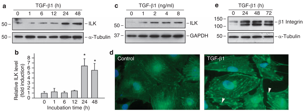
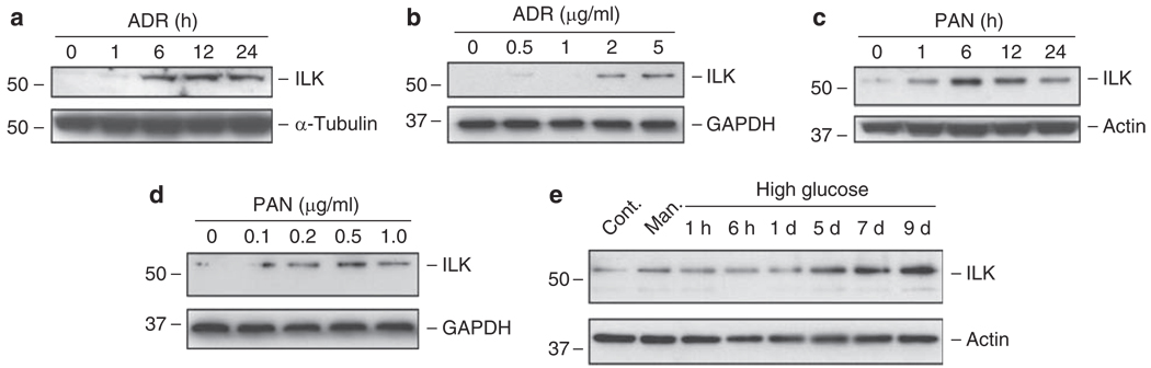
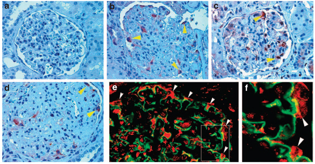
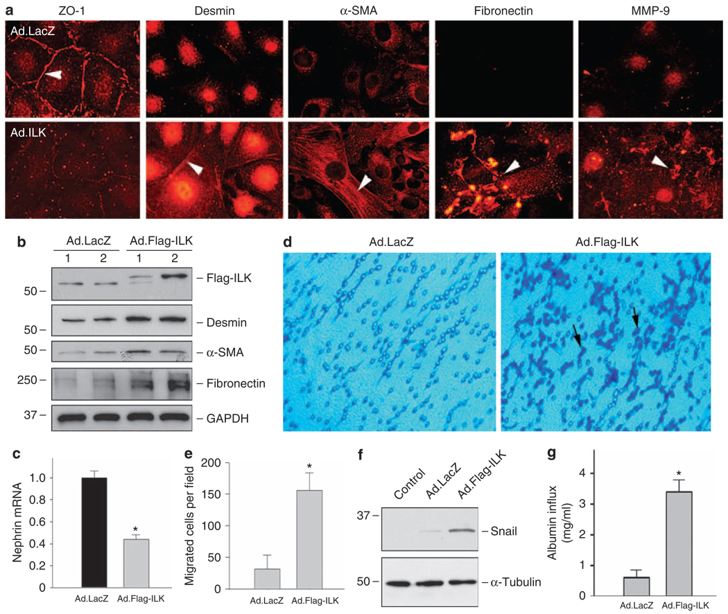
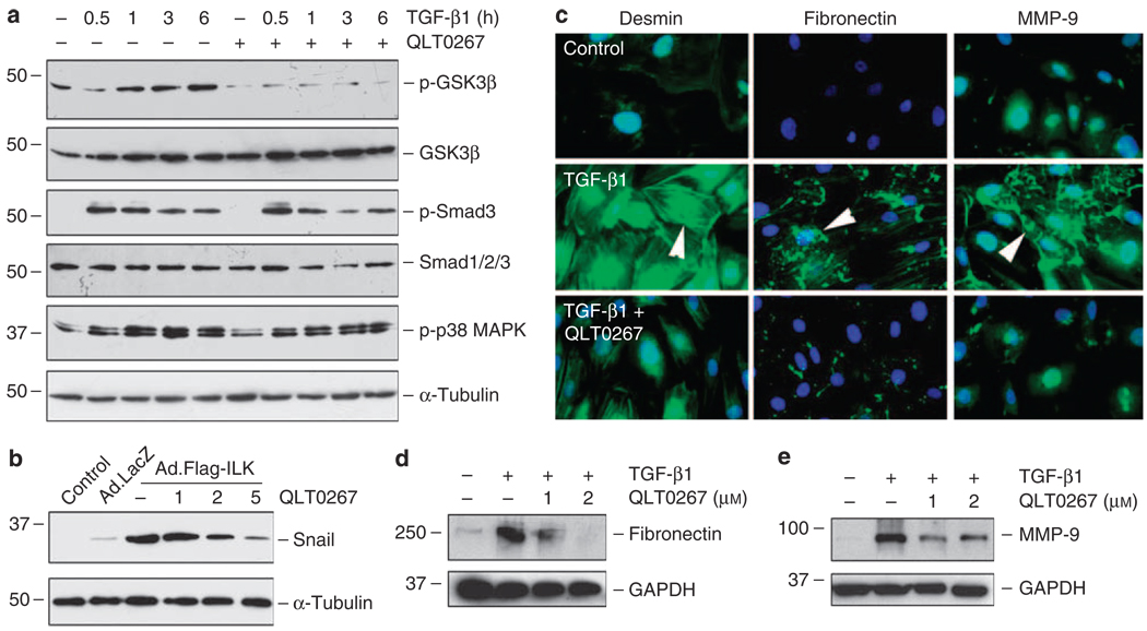
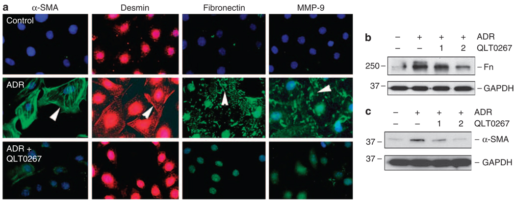
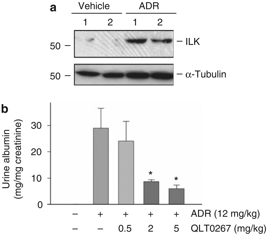
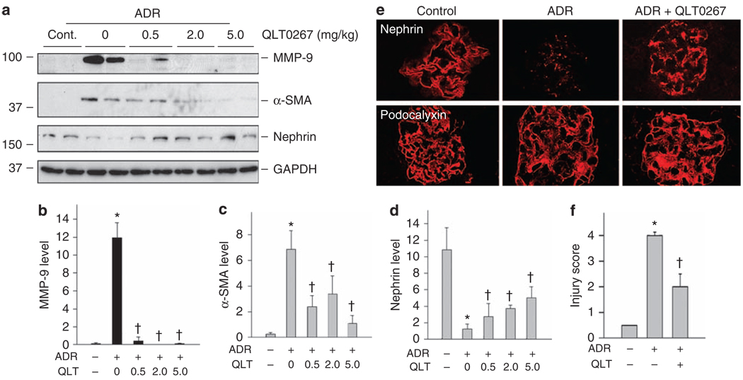
Proteinuria is a primary clinical symptom of a large number of glomerular diseases that progress to end-stage renal failure. Podocyte dysfunctions play a fundamental role in defective glomerular filtration in many common forms of proteinuric kidney disorders. Since binding of these cells to the basement membrane is mediated by integrins, we determined the role of integrin-linked kinase (ILK) in podocyte dysfunction and proteinuria. ILK expression was induced in mouse podocytes by various injurious stimuli known to cause proteinuria including TGF-beta1, adriamycin, puromycin, and high ambient glucose. Podocyte ILK was also found to be upregulated in human proteinuric glomerular diseases. Ectopic expression of ILK in podocytes decreased levels of the epithelial markers nephrin and ZO-1, induced mesenchymal markers such as desmin, fibronectin, matrix metalloproteinase-9 (MMP-9), and alpha-smooth muscle actin (alpha-SMA), promoted cell migration, and increased the paracellular albumin flux across podocyte monolayers. ILK also induced Snail, a key transcription factor mediating epithelial-mesenchymal transition (EMT). Blockade of ILK activity with a highly selective small molecule inhibitor reduced Snail induction and preserved podocyte phenotypes following TGF-beta1 or adriamycin stimulation. In vivo, this ILK inhibitor ameliorated albuminuria, repressed glomerular induction of MMP-9 and alpha-SMA, and preserved nephrin expression in murine adriamycin nephropathy. Our results show that upregulation of ILK is a convergent pathway leading to podocyte EMT, migration, and dysfunction. ILK may be an attractive target for therapeutic intervention of proteinuric kidney diseases.


Figures

Figure 1
Figure 1. TGF-β1 induces ILK and β1 integrin expression in mouse podocytes
(ac) Western blot analyses show that TGF-β1 induced ILK expression in cultured podocytes in a time- and dose-dependent manner. Podocytes were incubated with 2 ng/ml of TGF-β1 for various periods of time as indicated (a, b) or with different concentrations of TGF-β1 for 48 h (c). Quantitative determination of relative ILK levels in different times after TGF-β1 treatment was presented in (b). *P < 0.05 versus controls (n = 3). (d) Immunofluorescence staining showed ILK induction by TGF-β1 in podocytes. ILK protein was specifically localized at the focal adhesion sites of podocytes (arrowheads). (e) TGF-β1 (2 ng/ml) also induced β1 integrin expression in mouse podocytes. GAPDH, glyceraldehyde 3-phosphate dehydrogenase; ILK, integrin-linked kinase; TGF-β1, transforming growth factor-β1.
Figure 2
Figure 2. Induction of ILK expression in podocytes in response to various injurious stimuli
Podocytes were treated with 2 µg/ml of adriamycin (a) or with 1 µg/ml of puromycin aminonucleoside (c) for various periods of time as indicated, or with increasing amounts of ADR (b) and PAN (d) for 12 h. Podocytes were also incubated with high glucose (30 mm) for different periods of time as indicated (e). Both normal glucose (5 mm) and mannitol (5 mm normal glucose and 25 mm mannitol) were used as controls and harvested at 9 days. Cell lysates were immunoblotted with antibodies against ILK and housekeeping proteins. ADR, adriamycin; Cont. Man., control mannitol; GAPDH, glyceraldehyde 3-phosphate dehydrogenase; ILK, integrin-linked kinase; PAN, puromycin aminonucleoside.
Figure 3
Figure 3. Induction of ILK protein in the glomerular podocytes in patients with proteinuric kidney diseases
Immunohistochemical staining showed strong ILK expression in the glomerular podocytes of patients with proteinuric kidney diseases. (a) normal kidney; (b) focal segmental glomerulosclerosis; (c) focal segmental glomerulosclerosis; (d) diabetic nephropathy. (e) Double immunofluorescence staining shows ILK localization in glomerular podocytes. Kidney biopsies from patients with diabetic nephropathy were double immunostained for ILK (red) and podocyte marker synaptopodin (green). (f) Enlarged box area in (e). Arrowheads indicate positive staining.
Figure 4
Figure 4. Ectopic expression of ILK induces podocyte EMT, migration, and Snail expression
(a) Immunofluorescence staining showed that exogenous ILK expression resulted in suppression of ZO-1 and induction of mesenchymal marker desmin, α-SMA, fibronectin, and MMP-9 in podocytes. Podocytes were infected with adenovirus harboring either Flag-ILK (Ad.Flag-ILK) or β-galactosidase gene (Ad.LacZ). Overexpression of exogenous ILK (Flag-ILK) and induction of desmin, α-SMA and fibronectin in podocytes were also confirmed by western blot analysis (b). Of note, infection with control adenovirus (Ad.LacZ) did not significantly affect the basal expression of ILK, desmin, α-SMA and fibronectin. (c) Real-time RT-PCR showed that ectopic expression of exogenous ILK inhibited nephrin mRNA expression. Relative mRNA level over the Ad.LacZ controls (value = 1.0) is presented. *P < 0.05 (n = 6). (d, e) Forced expression of ILK promoted podocyte migration. Representative micrographs show podocyte migration in a Boyden chamber motility assay (d). Quantitative determination of the migrated podocytes per field in different groups is presented (e). *P < 0.05 (n = 3). (f) Ectopic expression of ILK induced Snail expression. Cell lysates were prepared at 48 h after infection with Ad.Flag-ILK or Ad.LacZ adenovirus, and immunoblotted with antibodies against Snail and α-tubulin, respectively. Cells without infection with adenovirus were denoted as control. (g) Overexpression of ILK impaired the filtration barrier function of podocytes. Podocyte monolayer on collagen-coated Transwell filters was infected with Ad.LacZ or Ad.Flag-ILK adenovirus, and 24 h later albumin permeability across podocyte monolayer was determined. Data are presented as means ± s.e.m. (n = 6). *P < 0.01 versus Ad.LacZ control. α-SMA, α-smooth muscle actin; EMT, epithelial–mesenchymal transition, GAPDH, glyceraldehyde 3-phosphate dehydrogenase; ILK, integrin-linked kinase; MMP-9, matrix metalloproteinase-9.
Figure 5
Figure 5. Targeting ILK activity by small molecule inhibitor QLT0267 abolishes Snail induction and blocks mesenchymal conversion of podocytes
(a) Small molecule ILK inhibitor QLT0267 selectively inhibited the TGF-β1-induced phosphorylation of GSK-3β, but not Smad3 and p38 MAPK. Podocytes were pretreated with QLT0267 (10 µm) for 0.5 h, followed by incubation with TGF-β1 (2 ng/ml) for different periods of time as indicated. Cell lysates were immunoblotted with various antibodies as indicated. (b) QLT0267 blocked ILK-induced Snail expression in podocytes. Podocytes were infected with Ad.Flag-ILK or Ad.LacZ adenovirus. At 24 h later, cells were treated with different doses of QLT0267 as indicated for additional 24 h. Cell lysates were immunoblotted with specific antibodies against Snail and α-tubulin. (c) Immunofluorescence staining showed that QLT0267 inhibited the desmin, fibronectin, and MMP-9 expression induced by TGF-β1 in podocytes. (d, e) Western blot analyses showed that QLT0267 inhibited the TGF-β1-mediated fibronectin and MMP-9 expression in a dose-dependent manner. Podocytes were treated without or with TGF-β1 (2 ng/ml) in the absence or presence of different doses of QLT0267 as indicated. GAPDH, glyceraldehyde 3-phosphate dehydrogenase; GSK-3β, glycogen synthase kinase-3β; ILK, integrin-linked kinase; MAPK, mitogen-activated protein kinase; MMP-9, matrix metalloproteinase-9; TGF-β1, transforming growth factor-β1.
Figure 6
Figure 6. QLT0267 also blocks the ADR-mediated mesenchymal conversion of podocytes
(a) Immunofluorescence staining showed that QLT0267 inhibited the α-SMA, desmin, fibronectin, and MMP-9 expression induced by ADR in podocytes. (b, c) Western blot analyses showed that QLT0267 dose dependently inhibited the ADR-mediated fibronectin and α-SMA expression in podocytes. Doses of QLT0267 (1 and 2 µm) are indicated. ADR, adriamycin; α-SMA, α-smooth muscle actin, Fn, fibronectin; GAPDH, glyceraldehyde 3-phosphate dehydrogenase.
Figure 7
Figure 7. Targeting ILK in vivo ameliorates albuminuria after ADR injury
(a) Upregulation of ILK in the glomeruli in vivo after podocyte injury induced by ADR. Glomeruli were isolated from mice injected with ADR for 7 days. Whole-glomerular lysates were immunoblotted with antibodies against ILK and α-tubulin. Numbers (1 and 2) indicate each individual animal in a given group. (b) Inhibition of ILK activity by QLT0267 reduced albuminuria in ADR nephropathy. Mice injected with ADR were administrated with different doses of QLT0267 for 7 days. Urinary albumin level was determined and corrected to urine creatinine. Urine albumin was expressed as mg per mg creatinine and presented as mean ± s.e.m. *P < 0.05 versus ADR alone (n = 6). ADR, adriamycin; ILK, integrin-linked kinase.
Figure 8
Figure 8. Targeting ILK in vivo ameliorates the ADR-mediated glomerular injury and preserves nephrin expression
(ad) Whole-glomerular lysates were analyzed by western blotting using specific antibodies against MMP-9, α-SMA, nephrin, and GAPDH. (a) Representative western blots. (bd) Quantitative data on MMP-9 (b), α-SMA (c), and nephrin (d) are presented as mean ± s.e.m. (n = 6). *P < 0.05 versus normal controls, P < 0.05 versus ADR alone controls. (e) Confocal immunofluorescence microscopy showed the distribution of nephrin and podocalyxin in mouse glomeruli at 7 days after ADR injection. QLT0267 largely preserved nephrin expression after ADR. (f) Semiquantitative evaluation on nephrin loss (injury score) in the glomeruli among different groups. *P < 0.05 versus normal controls, P < 0.05 versus ADR alone (n = 6). ADR, adriamycin; α-SMA, α-smooth muscle actin; GAPDH, glyceraldehyde 3-phosphate dehydrogenase; ILK, integrin-linked kinase; MMP-9, matrix metalloproteinase-9.

References

    1. Shankland SJ. The podocyte’s response to injury: role in proteinuria and glomerulosclerosis. Kidney Int. 2006;69:2131–2147. - PubMed
    1. Wiggins RC. The spectrum of podocytopathies: a unifying view of glomerular diseases. Kidney Int. 2007;71:1205–1214. - PubMed
    1. Pavenstadt H, Kriz W, Kretzler M. Cell biology of the glomerular podocyte. Physiol Rev. 2003;83:253–307. - PubMed
    1. Tryggvason K, Wartiovaara J. Molecular basis of glomerular permselectivity. Curr Opin Nephrol Hypertens. 2001;10:543–549. - PubMed
    1. Reiser J, Kriz W, Kretzler M, et al. The glomerular slit diaphragm is a modified adherens junction. J Am Soc Nephrol. 2000;11:1–8. - PubMed

Publication types

MeSH terms