. 2010 May 27;465(7297):492-6.
doi: 10.1038/nature09075.

CD95 promotes tumour growth

Affiliations

CD95 promotes tumour growth

Lina Chen et al. Nature. .

Erratum in

  • Nature. 2011 Jul 14;475(7355):254
  • Nature. 2011 Mar 10;471(7337):254
  • Nature. 2012 Nov 29;491(7426):784

Abstract

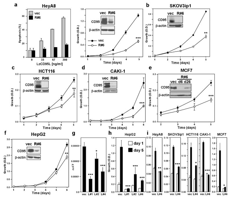
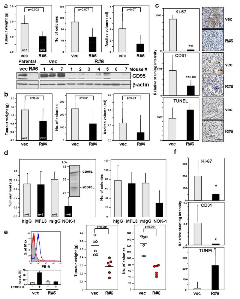
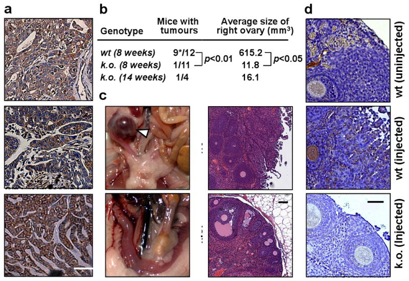
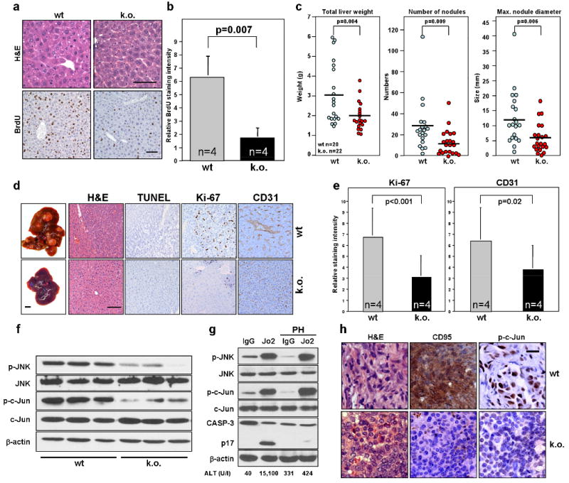
CD95 (also called Fas and APO-1) is a prototypical death receptor that regulates tissue homeostasis mainly in the immune system through the induction of apoptosis. During cancer progression CD95 is frequently downregulated or cells are rendered apoptosis resistant, raising the possibility that loss of CD95 is part of a mechanism for tumour evasion. However, complete loss of CD95 is rarely seen in human cancers and many cancer cells express large quantities of CD95 and are highly sensitive to CD95-mediated apoptosis in vitro. Furthermore, cancer patients frequently have elevated levels of the physiological ligand for CD95, CD95L. These data raise the possibility that CD95 could actually promote the growth of tumours through its non-apoptotic activities. Here we show that cancer cells in general, regardless of their CD95 apoptosis sensitivity, depend on constitutive activity of CD95, stimulated by cancer-produced CD95L, for optimal growth. Consistently, loss of CD95 in mouse models of ovarian cancer and liver cancer reduces cancer incidence as well as the size of the tumours. The tumorigenic activity of CD95 is mediated by a pathway involving JNK and Jun. These results demonstrate that CD95 has a growth-promoting role during tumorigenesis and indicate that efforts to inhibit its activity rather than to enhance it should be considered during cancer therapy.


Figures

Figure 1
Figure 1. Reducing CD95 or CD95L expression inhibits cell proliferation of cancer cells
a-f, Growth of different cell lines infected with the CD95 specific shRNA lentivirus CD95shRNA#6 (R#6). Inserts show the total CD95 expression levels of cells expressing scrambled control (vec) or R#6 as determined by western blot analysis. A similar effect was also found with a CD95 expressing variant of the neuroblastoma cell line NB4 (not shown). Apoptosis sensitivity of HeyA8 vec and R#6 cells by LzCD95 ligand treatment was determined by quantifying DNA fragmentation (a). g, HepG2 cells were stably infected with three different CD95L specific shRNA lentiviruses and CD95L mRNA was quantified using real time PCR. h, Growth of cells in g over 5 days. i, Growth of different cell lines infected with the L#4 virus. Proliferation of cells was examined by SRB assay (a-f and h and i). * p<0.05, ** p<0.01, *** p<0.001. Values in graphs in a to i represent mean -/+ s.d. from three independent experiments.
Figure 2
Figure 2. Loss of CD95 expression inhibits ovarian cancer in vivo
a Tumour weight, number of tumour colonies and ascites from mice injected with SKOV3ip1 vec or R#6 cells. Lysates of cells and tumour tissues were examined for CD95 level by western blot analysis. b, Same parameters as in (a) were measured from mice injected with SKOV3ip1 vec or R#4 cells. c, Histology and immunohistochemistry staining for Ki-67, TUNEL and CD31 of SKOV3ip1 vec and R#6 tumours. Scale bar = 100 μm. ** p-value <0.001. d, Tumour load and number of tumour colonies of mice treated with neutralising mAb for murine CD95L (MFL3), human CD95L (NOK-1) or corresponding isotype control mAbs were measured. Inset, Western blot analysis of SKOV3ip1 cell lysate for CD95L. e, Surface CD95 staining (upper left) and apoptosis sensitivity by LzCD95L (100 ng/ml) treatment (lower left) of MONTY-1 vec and R#6 cells. Weight and number of colonies of tumours formed by MONTY-1 vec and R#6 cells are shown (right). f, The staining intensity for Ki-67, TUNEL and CD31 of tumours from MONTY-1 vec and R#6 cells were quantified. * p-value <0.05. Values in graphs in a to e and f represent mean -/+ s.d. from three independent experiments. The horizontal bars in right part part of e represent the mean of 6 animals.
Figure 3
Figure 3. Deletion of CD95 leads to reduction in tumour formation in a spontaneous model of endometrioid ovarian cancer
a, Staining for CD95 in three primary endometrioid ovarian cancers. Scale bar = 50 μm. b, Number of mice that formed visible tumours either 8 or 14 weeks after injection of AdV Cre into the right ovarian bursa. * one mouse died from ovarian cancer 42 days after AdV Cre injection. c, Representative image and histology of right ovaries from wt and k.o. mice 8 weeks after injection of AdV Cre. Arrow head indicates ovarian tumour. Scale bar = 100 μm. d, CD95 staining of ovary from untreated wt, AdV Cre treated wt or AdV Cre treated k.o. mice. Scale bar = 50 μm. wt, LSL-K-rasG12D/+PtenloxP/loxPCD95wt/wt; k.o., K-rasG12D/+PtenloxP/loxPCD95loxP/loxP.
Figure 4
Figure 4. Deletion of CD95 in the liver leads to a decrease in tumour formation caused by reduced ability of hepatocytes to proliferate and to activate JNK
a, H&E and BrdU staining of livers from wild-type (wt) and liver specific CD95 k.o. mice 48 hrs after partial hepatectomy. Scale bars = 100 μm. b, Quantification of relative BrdU staining intensity of the mice in (a). c, wt and liver specific CD95 k.o. mice were injected with a single dose of DEN i.p. to induce liver tumour formation. 8 months later, all mice were sacrificed and parameters of total liver weight, number of liver surface nodules and maximum nodule diameter were recorded. d, Intact livers (scale bar = 1 cm), H&E staining and immunohistochemistry of TUNEL, Ki-67 and CD31 from mice in (c). Scale bar = 100 μm. e, Quantification of Ki-67 and CD31 staining for liver samples in (d). There was no detectable TUNEL staining. f, Western blot for phospho-JNK and phospho-c-Jun in three untreated wt and three CD95 k.o. mouse livers. g, wt mice with or without PH were injected i.p with 10 μg of murine CD95-specific agonistic antibody, Jo2 or isotype matched control mAb. After 6 hours phospho-JNK, p-c-Jun levels and cleavage of caspase-3 in livers were measured by western blot analysis. Concentration of the liver enzyme ALT in the serum of injected mice is given. h, Immunohistochemistry staining of ovarian tumours from wt (LSL-K-rasG12D/+PtenloxP/loxPCD95wt/wt) or k.o. (K-rasG12D/+PtenloxP/loxPCD95loxP/loxP) mice 8 weeks after injection of Adv Cre for CD95 and phospho-c-Jun. Scale bar = 20 μm. Values in graphs in b and e represent mean -/+ s.d. from three independent experiments. The horizontal bars in c represent the mean.

Comment in

References

    1. Nagata S. Fas ligand-induced apoptosis. Annu Rev Genet. 1999;33:29–55. - PubMed
    1. Krammer PH. CD95's deadly mission in the immune system. Nature. 2000;407:789–95. - PubMed
    1. Strasser A, Jost PJ, Nagata S. The many roles of FAS receptor signaling in the immune system. Immunity. 2009;30:180–92. - PMC - PubMed
    1. Peter ME, Legembre P, Barnhart BC. Does CD95 have tumor promoting activities? Biochim Biophys Acta. 2005;1755:25–36. - PubMed
    1. Debatin KM, Krammer PH. Death receptors in chemotherapy and cancer. Oncogene. 2004;23:2950–66. - PubMed

Publication types

MeSH terms