. 2010 May 21;5(5):e10755.
doi: 10.1371/journal.pone.0010755.

Eef1a2 promotes cell growth, inhibits apoptosis and activates JAK/STAT and AKT signaling in mouse plasmacytomas

Affiliations

Eef1a2 promotes cell growth, inhibits apoptosis and activates JAK/STAT and AKT signaling in mouse plasmacytomas

Zhaoyang Li et al. PLoS One. .

Abstract

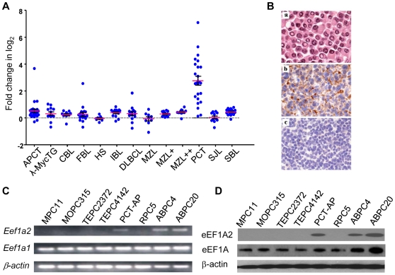
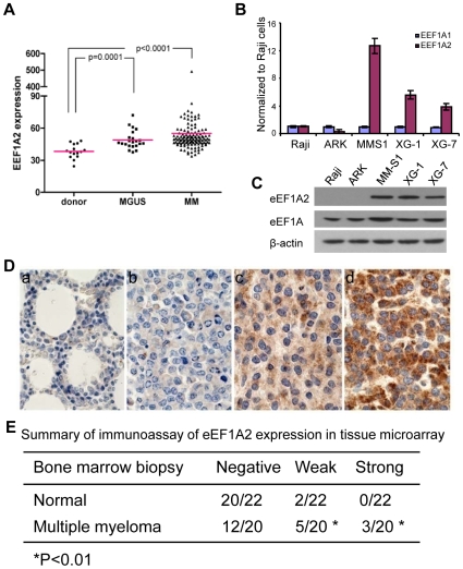
Background: The canonical function of EEF1A2, normally expressed only in muscle, brain, and heart, is in translational elongation, but recent studies suggest a non-canonical function as a proto-oncogene that is overexpressed in a variety of solid tumors including breast and ovary. Transcriptional profiling of a spectrum of primary mouse B cell lineage neoplasms showed that transcripts encoding EEF1A2 were uniquely overexpressed in plasmacytomas (PCT), tumors of mature plasma cells. Cases of human multiple myeloma expressed significantly higher levels of EEF1A2 transcripts than normal bone marrow plasma cells. High-level expression was also a feature of a subset of cell lines developed from mouse PCT and from the human MM.

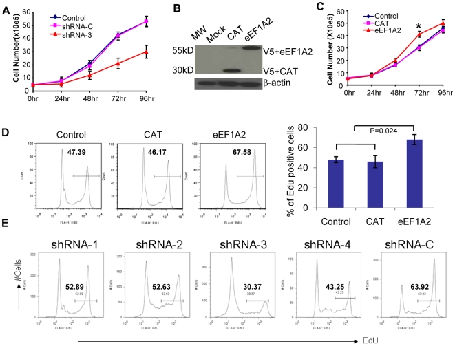
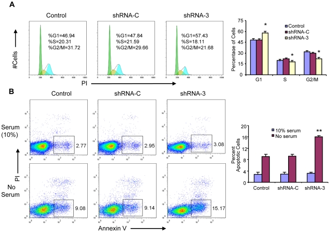
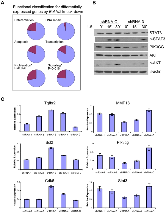
Methodology/principal findings: Heightened expression of EEF1A2 was not associated with increased copy number or coding sequence mutations. shRNA-mediated knockdown of Eef1a2 transcripts and protein was associated with growth inhibition due to delayed G1-S progression, and effects on apoptosis that were seen only under serum-starved conditions. Transcriptional profiles and western blot analyses of knockdown cells revealed impaired JAK/STAT and PI3K/AKT signaling suggesting their contributions to EEF1A2-mediated effects on PCT induction or progression.

Conclusions/significance: EEF1A2 may play contribute to the induction or progression of some PCT and a small percentage of MM. Eef1a2 could also prove to be a useful new marker for a subset of MM and, ultimately, a possible target for therapy.


Conflict of interest statement

Competing Interests: The authors have declared that no competing interests exist.

Figures

Figure 1
Figure 1. Expression of Eef1a2 in primary mouse lymphomas and PCT cell lines.
(A) Microarray analyses of Eef1a2 expression were performed among 11 classes of mouse primary B cell lymphomas and histiocytic sarcomas: APCT, anaplastic plasmacytoma ; λ-MycTG, lymphomas from λ-Myc transgenic mice ; CBL, centroblastic lymphoma ; FBL, follicular B cell lymphoma ; HS, histiocytic sarcoma ; IBL, immunoblastic lymphoma ; DLBCL, diffuse large B cell lymphoma ; MZL, marginal zone lymphoma (low grade); MZL+ and MZL++, marginal zone lymphoma (high grade) ; PCT, plasmacytoma ; SJL, SJL mouse lymphoma ; SBL, small B cell lymphoma . Each point represents a single tumor. The red line indicates the mean value for all tumors. The error bar  =  ± S.D. (B) Immunohistochemical studies of EEF1A2 expression using a polyclonal antibody specifically recognizing eEF1A2. a, H&E staining of PCT; b, IHC staining of PCT, c, IHC staining of MZL. (C, D) RT-PCR and Western blot analyses of Eef1a2 expression at the transcript (C) and protein (D) levels in PCT cell lines.
Figure 2
Figure 2. Expression of EEF1A2 in human MM and MM cell lines.
(A) Expression level of EEF1A2 in human MM determined by microarray. RNA from CD138+ plasma cells from the bone marrow of patients with MGUS, MM and donor, was hybridized to Affymetrix U133A microarrays. Data was from GEO (GSM6477). Unpaired t test with Welch's correction was carried out between MGUS or MM and donors. (B, C) RT-PCR and Western blot analyses of EEF1A2 expression at the transcript (B) and protein (C) levels in the indicated human MM cell lines. The error bar  =  ± S.D. (D) Immunohistochemistry analyses of EEF1A2 expression in normal costal tissue with negative staining (a) of EEF1A2 and MM tissues with negative staining (b), weak staining (c) and strong staining (d) of EEF1A2. (E) Summary of immunoassay of EEF1A2 expression in tissue microarray.
Figure 3
Figure 3. Knockdown of Eef1a2 inhibits cell growth and cell proliferation.
(A) ABPC4 cell numbers stably expressing Eef1a2 shRNA-3 and control shRNA-C cells were determined during four days in culture. (B, C) Overexpression of EEF1A2 protein in the MPC11 cell line (B). Cell numbers were determined during culture after transfection (C). (D) The frequency of EdU-postive MPC11 cells in transiently transfected Eef1a2 and control plasmid was analyzed by flow cytometry (left). The statistic bars show on the right. (E) The frequency of EdU-postive PCT-AP cells in transiently transfected Eef1a2 shRNAs, and control shRNA expressing PCT-AP cells was analyzed by flow cytometry.
Figure 4
Figure 4. Eef1a2 knockdown delays cell cycle entry and increases apoptosis induced by serum-free medium.
(A) Cell cycle analyses of PI-stained Eef1a2 shRNA and control cells using flow cytometry. (B) Early stage apoptotic cells were analyzed by flow cytometry in stably expressing Eef1a2 and control shRNAs and control cells after culturing in serum-free medium for 12 hours. Error bar  =  ± S.E. *p<0.05, **p<0.01.
Figure 5
Figure 5. Functional changes after Eef1a2 knockdown.
(A) Functional classification of differentially expressed genes by Eef1a2 knockdown cells. (B) Downregulation of Eef1a2 impaired IL-6-induced AKT and STAT3 phosphorylation. Eef1a2 shRNA and control cells were treated with 100 ng/ml recombinant IL-6 and protein samples were prepared 15 and 30 min later. Western blot analyses were performed using the indicated antibodies. (C) qPCR analyses of gene expression levels in transiently transfected shRNAs in PCT-AP cell line with four plasmids expressing specifically targeting Eef1a2 (shRNA-1,2,3,4) and a control plasmid (shRNA-C). Error bar  =  ± S.E.

Similar articles

Cited by

References

    1. Tomlinson VA, Newbery HJ, Wray NR, Jackson J, Larionov A, et al. Translation elongation factor eEF1A2 is a potential oncoprotein that is overexpressed in two-thirds of breast tumours. BMC Cancer. 2005;5:113. - PMC - PubMed
    1. Li R, Wang H, Bekele BN, Yin Z, Caraway NP, et al. Identification of putative oncogenes in lung adenocarcinoma by a comprehensive functional genomic approach. Oncogene. 2006;25:2628–2635. - PubMed
    1. Kulkarni G, Turbin DA, Amiri A, Jeganathan S, Andrade-Navarro MA, et al. Expression of protein elongation factor eEF1A2 predicts favorable outcome in breast cancer. Breast Cancer Res Treat. 2007;102:31–41. - PubMed
    1. Grassi G, Scaggiante B, Farra R, Dapas B, Agostini F, et al. The expression levels of the translational factors eEF1A 1/2 correlate with cell growth but not apoptosis in hepatocellular carcinoma cell lines with different differentiation grade. Biochimie. 2007;89:1544–1552. - PubMed
    1. Sun Y, Wong N, Guan Y, Salamanca CM, Cheng JC, et al. The eukaryotic translation elongation factor eEF1A2 induces neoplastic properties and mediates tumorigenic effects of ZNF217 in precursor cells of human ovarian carcinomas. Int J Cancer. 2008;123:1761–1769. - PMC - PubMed

Publication types

MeSH terms