. 2010 May 21;5(5):e10760.
doi: 10.1371/journal.pone.0010760.

UBIAD1 mutation alters a mitochondrial prenyltransferase to cause Schnyder corneal dystrophy

Affiliations

UBIAD1 mutation alters a mitochondrial prenyltransferase to cause Schnyder corneal dystrophy

Michael L Nickerson et al. PLoS One. .

Abstract

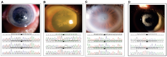
Background: Mutations in a novel gene, UBIAD1, were recently found to cause the autosomal dominant eye disease Schnyder corneal dystrophy (SCD). SCD is characterized by an abnormal deposition of cholesterol and phospholipids in the cornea resulting in progressive corneal opacification and visual loss. We characterized lesions in the UBIAD1 gene in new SCD families and examined protein homology, localization, and structure.

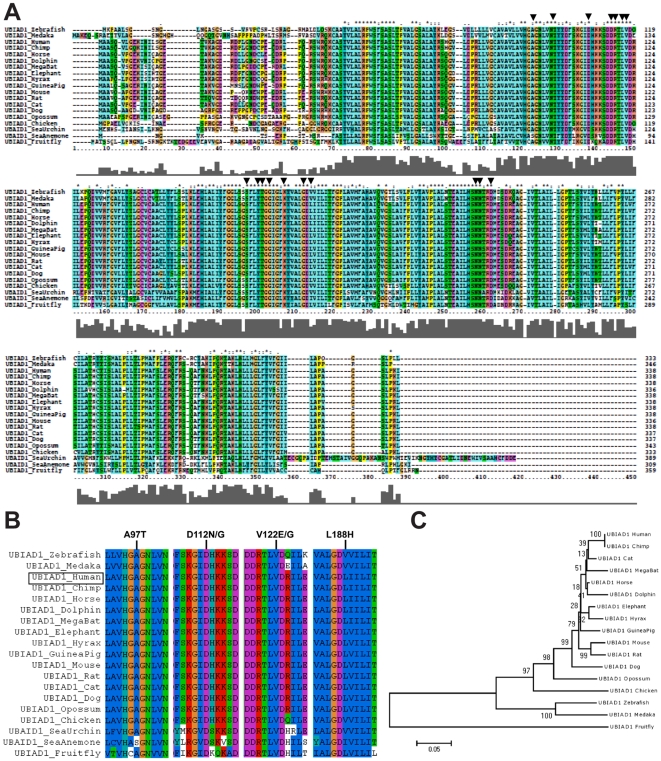
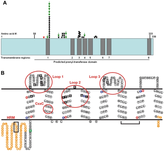
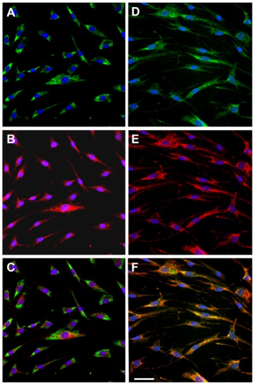
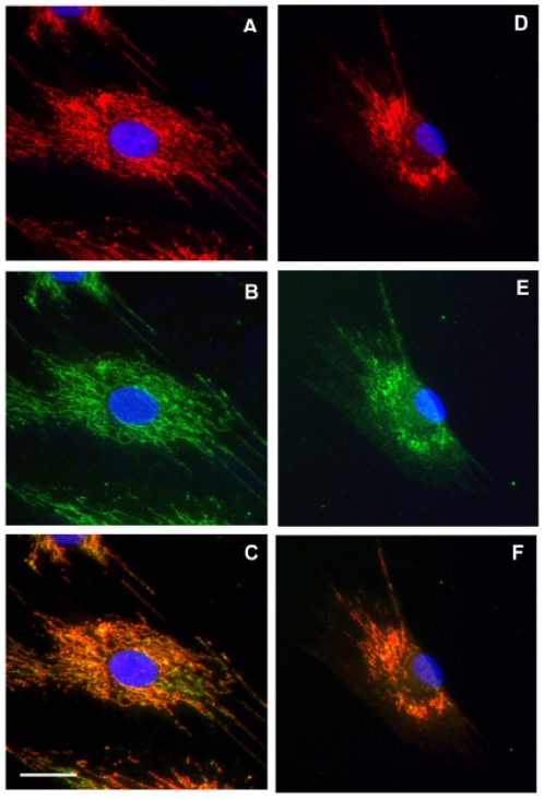
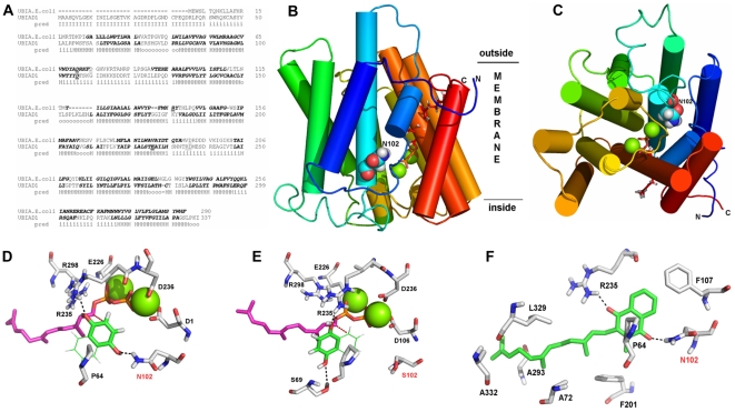
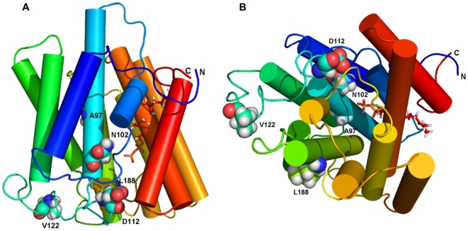
Methodology/principal findings: We characterized five novel mutations in the UBIAD1 gene in ten SCD families, including a first SCD family of Native American ethnicity. Examination of protein homology revealed that SCD altered amino acids which were highly conserved across species. Cell lines were established from patients including keratocytes obtained after corneal transplant surgery and lymphoblastoid cell lines from Epstein-Barr virus immortalized peripheral blood mononuclear cells. These were used to determine the subcellular localization of mutant and wild type protein, and to examine cholesterol metabolite ratios. Immunohistochemistry using antibodies specific for UBIAD1 protein in keratocytes revealed that both wild type and N102S protein were localized sub-cellularly to mitochondria. Analysis of cholesterol metabolites in patient cell line extracts showed no significant alteration in the presence of mutant protein indicating a potentially novel function of the UBIAD1 protein in cholesterol biochemistry. Molecular modeling was used to develop a model of human UBIAD1 protein in a membrane and revealed potentially critical roles for amino acids mutated in SCD. Potential primary and secondary substrate binding sites were identified and docking simulations indicated likely substrates including prenyl and phenolic molecules.

Conclusions/significance: Accumulating evidence from the SCD familial mutation spectrum, protein homology across species, and molecular modeling suggest that protein function is likely down-regulated by SCD mutations. Mitochondrial UBIAD1 protein appears to have a highly conserved function that, at least in humans, is involved in cholesterol metabolism in a novel manner.


Conflict of interest statement

Competing Interests: The authors have declared that no competing interests exist.

Figures

Figure 1
Figure 1. Corneas and UBIAD1 gene sequencing of SCD probands.
Corneal photos (top) and patient sequence chromatograms (bottom) are shown above a wild type sequence. (A) Proband from family GG with a novel A97T mutation. External photograph of the cornea demonstrating central and paracentral crystalline deposition in a 36 year old male. (B) Proband from family AA with a novel V122E mutation. The cornea shows central and paracentral crystalline deposits, diffuse corneal haze, and arcus lipoides in a 69 year old male (top). (C) Proband from family KK with a N102S mutation. The cornea shows central crystalline deposit, mid peripheral haze, and arcus lipoides in a 61 year old male. (D) Proband from family LL with a novel D112N mutation. The cornea shows paracentral crystalline deposition in a 25 year old male.
Figure 2
Figure 2. Highly conserved UBIAD1 residues are mutated in SCD.
(A) Locations of 17 amino acids mutated in SCD patients are indicated by arrows. Taller bars in the graph below the alignment indicate greater conservation. (B) Regions of alignment encompassing human SCD mutations: A97, D112, V122, L188, are shown. The position of the human protein in the alignment is indicated on the left (box). (C) Evolutionary relationships based upon UBIAD1 homology.
Figure 3
Figure 3. Locations of familial SCD alterations in UBIAD1.
(A) A linear diagram of the UBIAD1 protein with independent familial mutations (arrows). Novel familial mutations presented in this study are indicated by green arrows. Previously published SCD mutations are indicated (black arrows). Predicted transmembrane (grey boxes) and prenyltransferase domains (horizontal line, bottom) are indicated. A previously described S75F SNP is indicated (red arrow). Adapted from Ref. 11. (B) Locations of SCD mutations in a proposed 2-D model of UBIAD1 in a lipid bilayer. Solid Black: SCD mutations, Orange: amino acids outside the prenyltransferase domain, Blue: acidic residues, Red: basic residues, HRM: heme regulatory motif (box), CxxC: oxidoreductase motif (CAAC, small circle), Green: S75F polymorphism.
Figure 4
Figure 4. Cellular localization of wild type human UBIAD1.
Co-localization within cultured normal human keratocytes of UBIAD1 protein and protein disulfide isomerase, an enzyme in endoplasmic reticulum, is shown in panels A–C. Co-localization of UBIAD1 and OXPHOS complex I, an enzyme in mitochondria, is shown in D–F. UBIAD1 labeling is red (B and E). Protein disulfide isomerase and OXPHOS I are green (A and D). UBIAD1 did not co-localize with the endoplasmic reticulum (C), but did co-localize with mitochondria (co-localizing red and green show as orange in F). Bar is 50 µm and applies to all.
Figure 5
Figure 5. Localization of SCD mutant UBIAD1.
Co-localization of UBIAD1 and OXPHOS complex I mitochondrial marker in keratocytes derived from the Family KK proband (N102S mutation, panels A–C) and a healthy donor (D–F). UBIAD1 (red, A and D) and a mitochondrial marker (green, B and E) show co-localization (orange) in both normal (F) and SCD disease keratocytes (C). Bar is 25 µm and applies to all.
Figure 6
Figure 6. Three dimensional modeling of human UBIAD1.
(A) Alignment of E.coli UbiA and human UBIAD1 with predicted transmembrane helices (pred, bold italics), H = helix, I = inside, o = outside. Organic diphosphate binding residies are underlined. (B) Rainbow representation (side view) of a putative 3D-structure of UBIAD1 in the membrane. Approximate location of the lipid bilayer is indicated (horizontal lines). Inside and outside are arbitrary labels of membrane sidedness. Green spheres represent magnesium cations in the active site with a docked farnesyl-diphosphate (red stick representation). The side chain of N102 is shown as a space-filled atom. (C) Top view as described in Fig. 6B. Magenta atoms show potential binding of a putative substrate. (D) Hypothetical docking of farnesyldiphosphate and a 1,4-dihydroxy aryl compound. Substrate recognition by N102 (arrow) and R235 via hydrogen bonds and by hydrophobic interactions with P64 are indicated (dashed lines). The distance of the C2-atom of the hydroquinone to the C1-atom of the farnesyl moiety is 3.8 Å (red dashed line). (E) Docking arrangement of the two putative substrates as in Figure 6D upon in silico mutation of UBIAD1 from asparagine 102 to serine (arrow). The aromatic substrate is no longer recognized by N102, but by S69 and, as before, by R235 and P64. C2 of the aromatic substrate is no longer positioned correctly to allow prenylation. (F) Active site of UBIAD1 with a menaquinone-farnesyl derivative that optimally docks to the protein. Substrates with longer fatty acid tails were also successfully docked. The interaction is stabilized by hydrogen bonds (dashed lines) with N102 and R235. R235 may be influenced by neighboring residues, N232, N233, and D236, which cause SCD when altered. The quinone moiety and farnesyl chain are recognized by P64, F107, and other indicated residues via hydrophobic interactions.
Figure 7
Figure 7. Locations of selected SCD alterations.
(A) Sideview of UBIAD1 showing locations of wild type amino acids mutated in SCD. (B) Top view as in Figure 7A. In each view, only several residues mutated in SCD are visible. Farnesyldiphosphate is shown as a stick representation. The sidechains of SCD mutations reported in this paper are shown as spacefilled atoms: A97, N102, D112, V122, and L188.

References

    1. Van Went JM, Wibaut F. En Zeldzame erfelijk hoornvliessandoening. Niederl Tijdschr Geneesks. 1924;68:2996–2997.
    1. Schnyder WF. Mitteilung uber einen neuen typus von familiarer krankung. Schweiz Med Wschr. 1929;10:559–571.
    1. Rodrigues MM, Kruth H, Krachmer JH, Willis R. Unesterified cholesterol in Schnyder's corneal crystalline dystrophy. Am J Ophthalmol. 1987;104:157–163. - PubMed
    1. Bron AJ. Corneal changes in the dislipoproteinaemias. Cornea. 1989;8:75–79. - PubMed
    1. Weiss JS. Visual morbidity in thirty-four families with Schnyder Crystalline Corneal Dystrophy. Trans Am Ophthalmol Soc. 2007;105:616–648. - PMC - PubMed

Publication types

MeSH terms

LinkOut - more resources