. 2010 May 21;5(5):e10763.
doi: 10.1371/journal.pone.0010763.

Arginine metabolism by macrophages promotes cardiac and muscle fibrosis in mdx muscular dystrophy

Affiliations

Arginine metabolism by macrophages promotes cardiac and muscle fibrosis in mdx muscular dystrophy

Michelle Wehling-Henricks et al. PLoS One. .

Abstract

Background: Duchenne muscular dystrophy (DMD) is the most common, lethal disease of childhood. One of 3500 new-born males suffers from this universally-lethal disease. Other than the use of corticosteroids, little is available to affect the relentless progress of the disease, leading many families to use dietary supplements in hopes of reducing the progression or severity of muscle wasting. Arginine is commonly used as a dietary supplement and its use has been reported to have beneficial effects following short-term administration to mdx mice, a genetic model of DMD. However, the long-term effects of arginine supplementation are unknown. This lack of knowledge about the long-term effects of increased arginine metabolism is important because elevated arginine metabolism can increase tissue fibrosis, and increased fibrosis of skeletal muscles and the heart is an important and potentially life-threatening feature of DMD.

Methodology: We use both genetic and nutritional manipulations to test whether changes in arginase metabolism promote fibrosis and increase pathology in mdx mice. Our findings show that fibrotic lesions in mdx muscle are enriched with arginase-2-expressing macrophages and that muscle macrophages stimulated with cytokines that activate the M2 phenotype show elevated arginase activity and expression. We generated a line of arginase-2-null mutant mdx mice and found that the mutation reduced fibrosis in muscles of 18-month-old mdx mice, and reduced kyphosis that is attributable to muscle fibrosis. We also observed that dietary supplementation with arginine for 17-months increased mdx muscle fibrosis. In contrast, arginine-2 mutation did not reduce cardiac fibrosis or affect cardiac function assessed by echocardiography, although 17-months of dietary supplementation with arginine increased cardiac fibrosis. Long-term arginine treatments did not decrease matrix metalloproteinase-2 or -9 or increase the expression of utrophin, which have been reported as beneficial effects of short-term treatments.

Conclusions/significance: Our findings demonstrate that arginine metabolism by arginase promotes fibrosis of muscle in muscular dystrophy and contributes to kyphosis. Our findings also show that long-term, dietary supplementation with arginine exacerbates fibrosis of dystrophic heart and muscles. Thus, commonly-practiced dietary supplementation with arginine by DMD patients has potential risk for increasing pathology when performed for long periods, despite reports of benefits acquired with short-term supplementation.


Conflict of interest statement

Competing Interests: The authors have declared that no competing interests exist.

Figures

Figure 1
Figure 1. Mice with disrupted nNOS expression or localization do not exhibit inflammation or develop fibrosis.
(A and B) Cross-sections of 12-month old nNOS null (A) and α−sko (B) quadriceps stained with hematoxylin are free of inflammation. Bar = 50 µm. (C) Mononuclear cells in an inflammatory lesion of 18-month-old mdx quadriceps stain positive for arginase expression. Bar = 50 µm. (D) Mice lacking nNOS expression (nNOS ko) or localization to the sarcolemma (α-sko) do not develop pathological fibrosis in quadriceps (Quad), soleus, diaphragm (Diaph), longissimus dorsi (LD) and heart tissues. C57 mice (n = 5) and α-sko mice (n = 5) were 18-months old. nNOS ko mice (n = 5) were 12-months old.
Figure 2
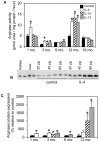
Figure 2. Th2 cytokines induce arginase activity and expression in mdx muscle macrophages.
(A) Arginase activity of muscle macrophages isolated from mdx mice at various ages was measured following in vitro stimulation with either IL-4, IL-10, IL-13 or no cytokine. At least 3 experiments were performed with a minimum of 5 wells for each age and condition. Some error bars are too small to be visible. (B) Representative western blot of 3-month mdx muscle macrophage lysates prepared following stimulation with IL-4, or no cytokine, loaded in various quantities as indicated and probed with an arginase-1 and 2 antibody. Homogenates of kidney, which expresses arginase-2, and liver, which expresses arginase-1, were included to show that the antibody recognizes both arginase isoforms. (C) Cytokines that induced increases in arginase activity increased arginase expression. Stimulated and control mdx muscle macrophages were analyzed by western blotting as in (B) and densitometrically quantified. * indicates statistical significance at p<0.05 as compared to age-matched control.
Figure 3
Figure 3. Arginase-2 mutation reduces muscle fibrosis.
(A) Mdx mice lacking arginase-2 expression demonstrated decreased fibrosis in quadriceps (Quad) and diaphragm (Diaph) muscles. Arginase-2 mutation reduced fibrosis in diaphragm, longissimus dorsi (LD) and heart of wild-type mice. Sol  =  soleus. n = 5 animals per group. (B-F) The concentration of macrophages, eosinophils, CD4+ cells, CD8+ cells and neutrophils in muscles were not affected by arginase-2 mutation. # indicates statistical significance at p<0.05 as compared to C57; ^ indicates statistical significance at p<0.05 as compared to mdx; * indicates statistical significance at p<0.05 as compared to A2ko/wt. n = 7 animals per group. (In A – F, some error bars are too small to be visible.) (G and H) Representative autoradiographs of C57 (G) and mdx (H) mice used to measure the kyphotic index (KI). The lines, AB and CD, used to calculate KI are indicated on (H). (I and J) Western blots for arginase-1 in wild-type, C57 mice (I) and mdx mice (J) show that null mutation of arginase-2 did not affect arginase-1 expression in muscles from either mouse line. All mice were 18-months old.
Figure 4
Figure 4. Absence of arginase-2 expression reduces collagen deposition.
Cross-sections of quadriceps from C57, arginase-2 null mice on a wild-type background (A2ko/wt), mdx and arginase-2 null mice on an mdx background (A2ko/mdx) were stained with antibodies specific for collagen type I (C1), collagen type 3 (C3), collagen type 4 (C4) and collagen type 5 (C5). No pathological fibrosis is evident in C57 or A2ko/wt quadriceps. Mdx mice develop fibrotic lesions and thickening of connective tissue which are reduced in A2ko/mdx mice. Bar = 50 µm. All mice were 18-months old.
Figure 5
Figure 5. Echocardiography shows that ablation of arginase-2 expression does not affect posterior wall thickness in the left ventricle of mdx or wild-type mice.
Image taken at the chordal level in the left ventricle shows relative motion of the interventricular septum (IVS) and posterior wall (PW) that delineate the end diastolic diameter (EDD) over time (left to right in image). Representative echocardiographs from (A) mdx (n = 5), (B) C57 (n = 7), (C) A2ko (n = 5) and (D) A2ko/mdx (n = 6) mice are shown. All mice were 18-months old.
Figure 6
Figure 6. Long-term supplementation with L-arginine increases fibrosis in dystrophin-deficient skeletal muscles and heart, without affecting inflammation.
(A) Connective tissue content of all dystrophin-deficient muscles assayed was significantly increased after 18 months of L-arginine treatment. There was no effect on C57 muscles. † indicates statistical significance at p<0.05 as compared to C57 samples. ^ indicates statistical significance at p<0.05 as compared to mdx mice treated with D-arginine as well as C57 samples. n = 5 animals per group. (B-F) Long-term treatment with L-arginine did not affect the muscle concentrations of macrophages, eosinophils, CD4+ cells, CD8+ cells or neutrophils. # indicates statistical significance at p<0.05 as compared to C57 D-arg samples. * indicates statistical significance at p<0.05 as compared to C57 L-arg samples. Quad  =  quadriceps, Sol  =  soleus, Diaph  =  diaphragm, LD  =  longissimus dorsi. n = 7 animals per group. Some error bars are too small to be visible.
Figure 7
Figure 7. Extended L-arginine treatment increases collagen deposition in mdx muscles.
Cross-sections of quadriceps isolated from C57 or mdx mice treated with L-arginine or D-arginine were labeled with antibodies to collagen type 1 (C1), collagen type 3 (C3), collagen type 4 (C4) and collagen 5 (C5). Collagen distribution is similar in C57 mice treated with D- or L-arginine (C57/D-arg and C57/L-arg, respectively). Significantly more collagen is present in mdx mice compared to C57 mice and collagen deposition is more prominent in mdx mice treated with L-arginine (mdx/L-arg) than mdx mice treated with D-arginine (mdx/D-arg). Bar = 50 µm. All mice were 18-months old.
Figure 8
Figure 8. Matrix metalloproteinases 2 and 9 were unaffected following L-arginine treatment.
(A) Gelatinase zymogram showing MMP-2 and –9 in muscle homogenates from long-term, L- or D-arginine-treated mdx or C57 mice. MMP-9 activity was detected at 100 kDa and MMP-2 activity was detected at 60 and 66 kDa. Human MMP-9 was used as a standard and migrates lower than mouse MMP-9 (47). (B) Densitometric analysis of cleared bands shows that MMP activity is greater in mdx tissues, but is not affected by L-arginine supplementation. * indicates significant difference at p<0.05 as compared to treatment-matched C57 samples. All mice were 18-months old.
Figure 9
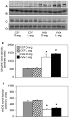
Figure 9. L-arginine treatment does not affect utrophin or nNOS expression.
Western blot of tibialis anterior muscle homogenates from long-term, L- or D-arginine-treated mdx or C57 mice probed for utrophin (A) or nNOS (C). Blots A and C were stripped and reprobed with anti-skeletal muscle alpha-actin (B and D, respectively) to verify loading consistency. Densitometry of protein expression assessed by western blotting shows that utrophin concentration is greater in mdx muscles but is unaffected by L-arginine treatment (E) and that nNOS concentration is greater in wild-type muscles but is unaffected by L-arginine treatment (F). * indicates statistical significance at p<0.05 as compared to treatment-matched C57 samples. All mice were 18-months old.
Figure 10
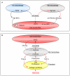
Figure 10. Model of potential, competitive interactions between mdx muscle fibers and macrophages for arginine in dystrophic muscles.
A: In 4-week muscles, M1 macrophages expressing iNOS and M2 macrophages expressing arginase are present in inflammatory lesions in mdx muscle (19). M1 macrophage iNOS and M2 macrophage arginase compete for their common substrate, arginine (19). In wild-type muscle, nNOS in muscle fibers also competes for arginine, but loss of nNOS from dystrophic muscle eliminates that competition, increasing arginine availability for iNOS, increasing cytolysis (19), or arginase, increasing fibrosis (present study). The numbers of iNOS expressing M1 macrophages decline after 4-weeks of age in mdx muscle increasing substrate availability for arginase, and the proportion of arginase-expressing macrophages increases by 12-months of age, further driving fibrosis (B). In addition to an increase in arginine for arginase, the loss of several negative regulatory influences of nNOS-derived NO fibrosis occurs. For example, nNOS-derived hydroxyarginine which can inhibit arginase would be diminished. Red arrows  =  profibrotic pathways. Black arrow  =  pathways that compete with fibrotic pathways. Broken arrow  =  pathways that are deficient in mdx muscle.

Similar articles

Cited by

References

    1. Ishizaki M, Suga T, Kimura E, Shiota T, Kawamo R, et al. Mdx respiratory impairment following fibrosis of the diaphragm. Neuromuscl Disord. 2008;18:342–348. - PubMed
    1. Inkley SR, Oldenburg FC, Vignos PJ. Pulmonary function in Duchenne muscular dystrophy related to stage of disease. Am J Med. 1974;56:297–306. - PubMed
    1. Moriuchi T, Kagawa N, Mukoyama M, Hizawa K. Autopsy analysis of the muscular dystrophies. Tokushima J Exp Med. 1993;40:83–93. - PubMed
    1. Rideau Y, Jankowski LW, Grellet J. Respiratory function in the muscular dystrophies. Muscle Nerve. 1981;4:155–164. - PubMed
    1. Laws N, Hoey A. Progression of kyphosis in mdx mice. J Appl Physiol. 2004;97:1970–1977. - PubMed

Publication types

MeSH terms