

Diffractive multifocal intraocular lens interferes with intraoperative view
- PMID: 20505840
- PMCID: PMC2874275
- DOI: 10.2147/opth.s8831
Diffractive multifocal intraocular lens interferes with intraoperative view
Abstract
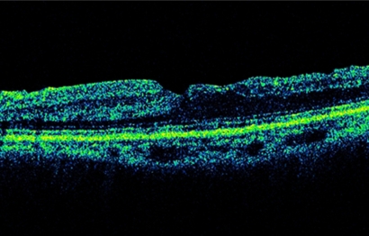
We report an unusual finding during vitreous surgery in an eye implanted with a diffractive multifocal intraocular lens (IOL). A 70-year-old woman reported gradual visual deterioration to 20/40 in the left eye two and a half years after uneventful cataract surgery with implantation of a diffractive multifocal IOL. Funduscopic examination showed an epiretinal membrane (ERM) in the left eye. Increased macular traction was believed to cause the visual deterioration. Vitreous surgery with removal of the ERM was performed and triamcinolone acetonide (TA) was injected intravitreally to visualize the residual vitreous cortex. Although the ERM was peeled successfully, the ability to focus on the vitreoretinal interface through the IOL required great effort with decreased contrast sensitivity and ghost images of the intravitreal TA crystals. The vision improved to 20/25 4 months postoperatively. Macular surgery can be performed in an eye with a diffractive multifocal IOL; however, decreased contrast sensitivity and ghost images may interfere with the intraoperative view through the diffractive IOL in complicated cases.
Keywords: diffractive multifocal intraocular lens; intraoperative view.
Figures

References
-
- Terwee T, Weeber H, van der Mooren M, et al. Visualization of the retinal image in eye model with spherical and aspheric, diffractive, and refractive multifocal intraocular lenses. J Refract Surg. 2008;24:223–232. - PubMed
-
- Charles S, Runge P. Vitreoretinal complications of multifocal intraocular lenses. In: Maxwell A, Nordan LT, editors. Multifocal Intraocular Lenses. Thorofare, NJ: Slack, Inc; 1991. pp. 209–218.
-
- Kumar A, Goyal M, Tewari HK. Posterior segment visualization problems with multifocal intraocular lenses. Acta Ophthalmol Scand. 1996;74:415. - PubMed
-
- Lim JI, Kuppermann BD, Gwon A, et al. Vitreoretinal surgery through multifocal intraocular lenses compared with monofocal intraocular lenses in fluid-filled and air-filled rabbit eyes. Ophthalmology. 2000;107:1083–1088. - PubMed
-
- Kawamura R, Inoue M, Shinoda K, et al. Intraoperative findings during vitreous surgery after implantation of diffractive multifocal intraocular lens. J Cat Ref Surg. 2008;34:1048–1049. - PubMed
Publication types
LinkOut - more resources
Full Text Sources
