. 2010 Aug;62(8):2382-91.
doi: 10.1002/art.27550.

Prevention of cartilage degeneration and restoration of chondroprotection by lubricin tribosupplementation in the rat following anterior cruciate ligament transection

Affiliations

Prevention of cartilage degeneration and restoration of chondroprotection by lubricin tribosupplementation in the rat following anterior cruciate ligament transection

Gregory D Jay et al. Arthritis Rheum. 2010 Aug.

Abstract

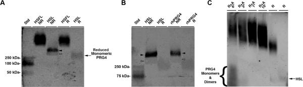
Objective: To investigate whether cartilage degeneration is prevented or minimized following intraarticular injections of lubricin derived from human synoviocytes in culture, recombinant human PRG4 (rhPRG4), or human synovial fluid (SF) in a rat model of anterior cruciate ligament (ACL) injury.

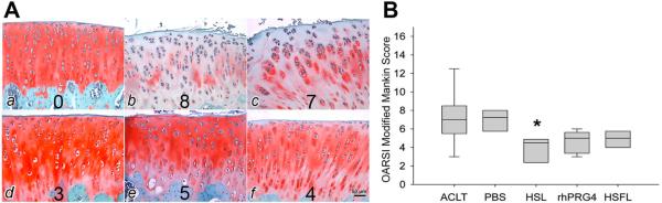
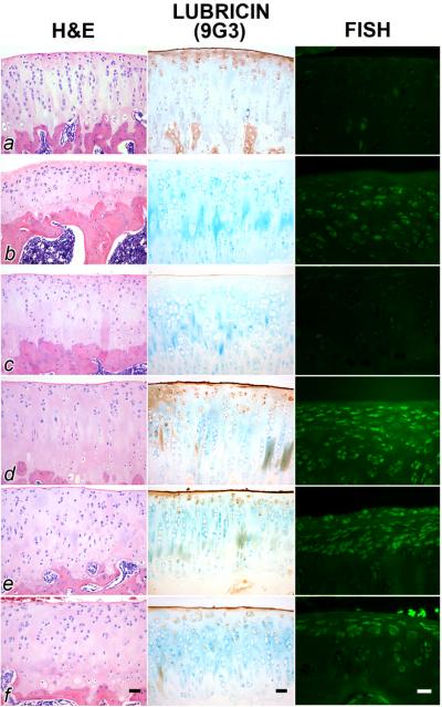
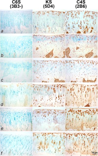
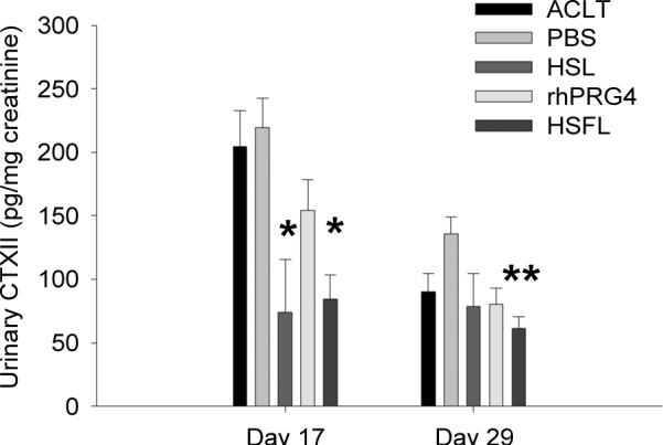
Methods: Unilateral ACL transection (ACLT) was performed in Lewis rats (n = 45). Nine animals were left untreated. The remaining rats were given intraarticular injections (50 microl/injection) of either phosphate buffered saline (PBS) (n = 9), human synoviocyte lubricin (200 microg/ml; n = 9), rhPRG4 (200 microg/ml; n = 9), or human SF lubricin (200 microg/ml; n = 9) twice weekly beginning on day 7 after injury. Joints were harvested on day 32 after injury. Histologic analysis was performed using Safranin O-fast green staining, and articular cartilage degeneration was graded using the Osteoarthritis Research Society International (OARSI)-modified Mankin criteria. Histologic specimens were immunoprobed for lubricin and sulfated glycosaminoglycans. A 24-hour urine collection was performed on days 17 and 29 postinjury, and urinary C-terminal telopeptide of type II collagen (CTX-II) levels were measured.

Results: Treatment with human synoviocyte lubricin resulted in significantly lower OARSI scores for cartilage degeneration compared with no treatment or PBS treatment (P < 0.05). Increased immunostaining for lubricin in the superficial zone chondrocytes and on the surface of cartilage was observed in lubricin-treated, but not untreated or PBS-treated, joints. On day 17, urinary CTX-II levels in human synoviocyte lubricin- and human SF lubricin-treated animals were significantly lower than those in untreated animals (P = 0.005 and P = 0.002, respectively) and in PBS-treated animals (P = 0.002 and P < 0.001, respectively).

Conclusion: After treatment with any of the 3 types of lubricin evaluated in this study, a reduction in cartilage damage following ACLT was evident, combined with a reduction in type II collagen degradation. Our findings indicate that intraarticular lubricin injection following an ACL injury may be beneficial in retarding the degeneration of cartilage and the development of posttraumatic OA.


Figures

Figure 1
Figure 1
Agarose gel electrophoresis of human synoviocyte lubricin (HSL), recombinant human proteoglycan 4 (rhPRG4), and human synovial fluid lubricin (HSFL) under reducing (R) and non-reducing (NR) conditions. A) Comparison of HSFL with HSL and B) HSL with rhPRG4. Dimerized HSL and rhPRG4 (solid arrowheads) and monomeric forms (open arrowheads) are reducible to a monomer with apparent MW 250 kDa. C) Progressive reduction and alkylation (R+A) measured in hours (°) or minutes (') of HSFL liberated monomeric and dimeric lubricin and resultant multimers assume a higher apparent molecular weight proportional to the duration of reduction and alkylation. The far right lane is HSL standard.
Figure 2
Figure 2
Safranin O stained tibial plateau cartilage and OARSI scores of control, untreated, PBS-treated, human synoviocyte lubricin (HSL), recombinant human proteoglycan 4 (rhPRG4), and human synovial fluid lubricin (HSFL). A. Representative tibial plateau cartilage from control (a), untreated (b), PBS-treated (c), HSL-treated (d), rhPRG4-treated (e), and HSFL-treated (f) animals harvested on day 32 post-ACLT. Associated consensus OASRI scores for these sections are illustrated at the bottom of each image. The tibial cartilage surface was rough along with inconsistent proteoglycan staining in untreated and PBS-treated joints, while the tibial cartilage surface was smooth, and proteoglycan staining was stronger in lubricin-treated joints. B. Box plot of OARSI modified Mankin scores of tibial plateaus from untreated (ACLT, n=9), PBS-treated (n=9), HSL-treated (n=9), rhPRG4-treated (n=9), and HSFL-treated (n=9) animals harvested on day 32 post-injury. Control cartilage was a criterion standard OARSI score = 0. *Indicates that HSL-treated joints had significantly lower (p<0.05) OARSI scores compared to ACLT and PBS-treated joints.
Figure 3
Figure 3
Hematoxylin and eosin (H&E) staining, mAb 9G3 immunostaining for lubricin and in situ hybridization for lubricin mRNA from ACL transected joints from articular cartilage from representative control (a), untreated (b), PBS-treated (c), human synoviocyte lubricin (HSL)-treated (d), recombinant human proteoglycan 4 (rhPRG4)-treated (e) and human synovial fluid lubricin (HSFL)-treated (f) animals. For joints treated with HSL (d), rhPRG4 (e) and HSFL (f), there was a greater surface accumulation of lubricin, and more superficial zone staining and lubricin expression. ACL-transected joints (b) and those treated with PBS (c) showed either no or minimal surface staining for lubricin. Scale bars are 20 μm.
Figure 4
Figure 4
Immunostaining of articular cartilage from ACL transected joints for chondroitin 6 sulfate (C6S), keratin sulfate (KS) and chondroitin 4 sulfate (C4S) from representative control (a), untreated (b), PBS-treated (c), human synoviocyte lubricin (HSL)-treated (d), recombinant human proteoglycan 4 (rhPRG4)-treated (e), and human synovial fluid lubricin (HSFL)-treated (f) animals. Controls and those treated with any lubricin generally showed more cellular staining for KS and C4S, than untreated or placebo-treated ACL transected joints. Greater matrix staining was noted for C4S across lubricin-treated joints. Joints treated with HSL (d) stained for C6S using 3B3 without chondroitinase digestion (3B3-) which may be indicative of repair activity.
Figure 5
Figure 5
Urinary CTXII (uCTXII) levels, normalized to creatinine, of untreated ACL transected joints (ACLT, n=6), PBS-treated (PBS, n=6), HSL-treated (HSL, n=3), rhPRG4 treated (rhPRG4, n=6) and HSFL-treated (HSFL, n=6) animals on days 17 and 29 post-injury. *Indicates that day 17 uCTXII levels of HSL and HSFL-treated animals were significantly lower than corresponding uCTXII levels of untreated (p=0.005; p=0.002) and PBS-treated (p=0.002; p<0.001) animals, respectively. **Indicates that day 29 uCTXII levels of HSFL-treated animals were significantly lower (p<0.001) than PBS-treated animals.

References

    1. Aichroth PM, Patel DV, Zorrilla P. The natural history and treatment of rupture of the anterior cruciate ligament in children and adolescents. A prospective review. J Bone Joint Surg Br. 2002;84(1):38–41. - PubMed
    1. Fithian DC, Paxton LW, Goltz DH. Fate of the anterior cruciate ligament-injured knee. Orthop Clin North Am. 2002;33(4):621–36. v. - PubMed
    1. Li G, Moses JM, Papannagari R, Pathare NP, DeFrate LE, Gill TJ. Anterior cruciate ligament deficiency alters the in vivo motion of the tibiofemoral cartilage contact points in both the anteroposterior and mediolateral directions. J Bone Joint Surg Am. 2006;88(8):1826–34. - PubMed
    1. Roos H, Adalberth T, Dahlberg L, Lohmander LS. Osteoarthritis of the knee after injury to the anterior cruciate ligament or meniscus: the influence of time and age. Osteoarthritis Cartilage. 1995;3(4):261–7. - PubMed
    1. Vasara AI, Jurvelin JS, Peterson L, Kiviranta I. Arthroscopic cartilage indentation and cartilage lesions of anterior cruciate ligament-deficient knees. Am J Sports Med. 2005;33(3):408–14. - PubMed

Publication types

MeSH terms