. 2010 Nov;225(2):569-76.
doi: 10.1002/jcp.22240.

The human SWI/SNF complex associates with RUNX1 to control transcription of hematopoietic target genes

Affiliations

The human SWI/SNF complex associates with RUNX1 to control transcription of hematopoietic target genes

Rachit Bakshi et al. J Cell Physiol. 2010 Nov.

Abstract

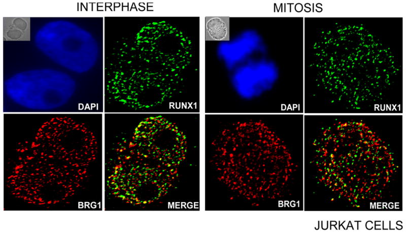
The acute myeloid leukemia 1 (AML1, RUNX1) transcription factor is a key regulator of hematopoietic differentiation that forms multi-protein complexes with co-regulatory proteins. These complexes are assembled at target gene promoters in nuclear microenvironments to mediate phenotypic gene expression and chromatin-related epigenetic modifications. Here, immunofluorescence microscopy and biochemical assays are used to show that RUNX1 associates with the human ATP-dependent SWI/SNF chromatin remodeling complex. The SWI/SNF subunits BRG1 and INI1 bind in vivo to RUNX1 target gene promoters (e.g., GMCSF, IL3, MCSF-R, MIP, and p21). These interactions correlate with histone modifications characteristic of active chromatin, including acetylated H4 and dimethylated H3 lysine 4. Downregulation of RUNX1 by RNA interference diminishes the binding of BRG1 and INI1 at selected target genes. Taken together, our findings indicate that RUNX1 interacts with the human SWI/SNF complex to control hematopoietic-specific gene expression.


Figures

Figure 1
Figure 1. Co-localization of RUNX1 and BRG1 proteins in Jurkat Cells
Immunofluorescence microscopy was performed to detect endogenous RUNX1 and BRG1 in Jurkat cells, and images were merged to determine co-localization. Immunofluorescence images were obtained using antibodies against RUNX1 (green) and BRG1 (red). The insets at top left of each panel show the phase contrast image. DAPI staining (blue) was used to visualize the nucleus (top row). RUNX1 and BRG1 proteins are predominantly present in the nucleus and display significant co-localization in interphase cells. Mitotic cells exhibit limited co-localization (less than 5% overlap in immunofluorescence signals).
Figure 2
Figure 2. Endogenous RUNX1 interacts with multiple subunits of the human SWI/SNF complex
A) Immunoprecipitation analysis was carried out with an antibody for RUNX1 in Jurkat cells. The precipitates were subjected to immunoblot analysis with BRG1, INI1, BAF155 and RUNX1 specific antibodies. All three proteins BRG1, INI1 and BAF155 were detected in western blot analysis as indicated in top three panels. B) Endogenous BRG1 and INI1 were immunoprecipitated from Jurkat cells using rabbit polyclonal antibodies. The precipitates were subjected to western blot analysis with RUNX1, BRG1 and INI1 antibodies. RUNX1 is present in immunoprecipitates obtained with either BRG1 or INI1 antibody.
Figure 3
Figure 3. RUNX1, BRG1 and INI1 associate with Runx target genes in interphase Jurkat cells
Chromatin immunoprecipitation was done with asynchronously growing Jurkat cells using RUNX1, BRG1, INI1 and IgG antibodies. Quantitative PCR was performed with specific primers from promoter regions of the following genes: CDK inhibitor p21 (CDKN1A), granulocyte-macrophage colony-stimulating factor (GMCSF), myeloperoxidase (MIP), interleukin 3 (IL-3), macrophage colony-stimulating factor 1 receptor (MCSF-R) and human osteocalcin (OC). The data show that endogenous RUNX1, BRG1 and INI1 bind to the promoters of the target genes in Jurkat cells. There is no significant binding of these proteins on the OC promoter, which is not expressed in these cells. Quantitative PCR data are normalized to genomic DNA and denoted as percent input.
Figure 4
Figure 4. RUNX1, BRG1 and INI1 occupancy of the GMCSF and IL3 promoters is associated with active histone modifications
Chromatin immunoprecipitations with RUNX1, BRG1, INI1, Pol II, IgG and histone modification antibodies were performed in Jurkat cells. The antibodies used to detect histone modifications were as follows: Acetylated histone H4 (H4-Ac), dimethylated H3 lysine 4 (H3K4me2) and dimethylated H3 lysine 27 (H3K27me2). The GMCSF and IL3 promoters are associated with dimethylated H3K4 and acetylated H4, but not dimethylated H3K27 (top panel). These two genes interact robustly with RNA polymerase II (lower panel), as well as with RUNX1, BRG1 and INI1 (middle panels). The human OC gene promoter is predominantly associated with dimethylated H3K27 and exhibits background binding for RNA polymerase II and the other factors. Quantitative PCR data are normalized to genomic DNA and denoted as percent input.
Figure 5
Figure 5. RUNX1 supports BRG1 and INI1 association with the GMCSF and IL3 promoters
Jurkat cells were transfected with two independent RUNX1 siRNAs and non-silencing (NS) Si-RNA control. A). To check the efficiency of knockdown, RUNX1 mRNA expression was measured by RT-qPCR relative to GAPDH in equal numbers of cells. B) Protein expression of RUNX1 was examined in siRNA treated samples as well as non silencing controls by western blot analysis using specific antibody. Immunoblotting with antibody against α-tubulin was performed as loading control. C). ChIP assays were performed in RUNX1 knockdown and control Jurkat cells with antibodies against RUNX1, BRG1, INI1 and IgG. Quantitative PCR data show that interactions of BRG1 and INI1 with the GMCSF and IL3 promoters are significantly reduced upon siRNA mediated RUNX1 knockdown. Quantitative PCR data are normalized to genomic DNA and denoted as percent input. D). Expression levels of GMCSF and IL3 mRNAs were analyzed by real-time quantitative RT-PCR relative to GAPDH in control and siRNA treated samples. Decreased association of RUNX1, BRG1 and INI1 to the GMCSF and IL3 promoters correlates with a reduced expression of these genes as measured by qRT-PCR.

References

    1. Armstrong JA, Bieker JJ, Emerson BM. A SWI/SNF-related chromatin remodeling complex, E-RC1, is required for tissue-specific transcriptional regulation by EKLF in vitro. Cell. 1998;95:93–104. - PubMed
    1. Bernardin-Fried F, Kummalue T, Leijen S, Collector MI, Ravid K, Friedman AD. AML1/RUNX1 increases during G1 to S cell cycle progression independent of cytokine-dependent phosphorylation and induces cyclin D3 gene expression. J Biol Chem. 2004;279:15678–15687. - PubMed
    1. Biggs JR, Peterson LF, Zhang Y, Kraft AS, Zhang DE. AML1/RUNX1 phosphorylation by cyclin-dependent kinases regulates the degradation of AML1/RUNX1 by the anaphase-promoting complex. Mol Cell Biol. 2006;26:7420–7429. - PMC - PubMed
    1. Bochar DA, Wang L, Beniya H, Kinev A, Xue Y, Lane WS, Wang W, Kashanchi F, Shiekhattar R. BRCA1 is associated with a human SWI/SNF-related complex: linking chromatin remodeling to breast cancer. Cell. 2000;102:257–265. - PubMed
    1. Brown SA, Imbalzano AN, Kingston RE. Activator-dependent regulation of transcriptional pausing on nucleosomal templates. Genes Dev. 1996;10:1479–1490. - PubMed

Publication types

MeSH terms

LinkOut - more resources