. 2010 Oct 1;111(2):284-94.
doi: 10.1002/jcb.22694.

Osteoblast lineage cells expressing high levels of Runx2 enhance hematopoietic progenitor cell proliferation and function

Affiliations

Osteoblast lineage cells expressing high levels of Runx2 enhance hematopoietic progenitor cell proliferation and function

Brahmananda R Chitteti et al. J Cell Biochem. .

Abstract

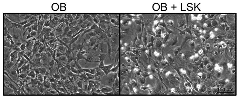
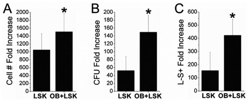
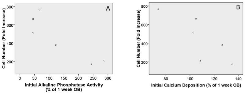
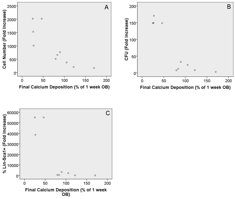
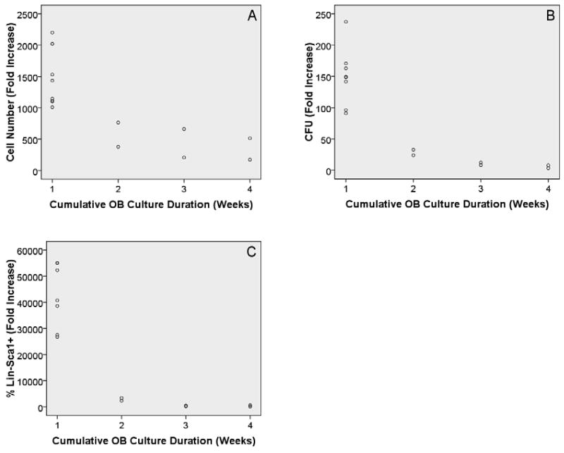
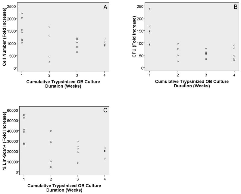
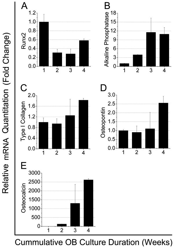
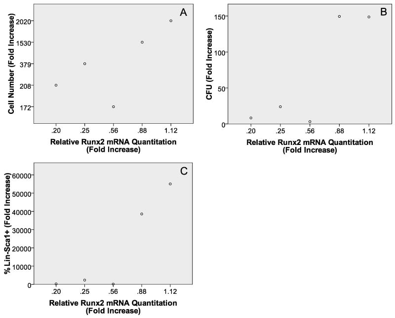
Although osteoblasts (OB) play a key role in the hematopoietic stem cell (HSC) niche, little is known as to which specific OB lineage cells are critical for the enhancement of stem and progenitor cell function. Unlike hematopoietic cells, OB cell surface phenotypic definitions are not well developed. Therefore, to determine which OB lineage cells are most important for hematopoietic progenitor cell (HPC) function, we characterized OB differentiation by gene expression and OB function, and determined whether associations existed between OB and HPC properties. OB were harvested from murine calvariae, used immediately (fresh OB) or cultured for 1, 2, or 3 weeks prior to their co-culture with Lin(-)Sca1(+)c-kit(+) (LSK) cells for 1 week. OB gene expression, alkaline phosphatase activity, calcium deposition, hematopoietic cell number fold increase, CFU fold increase, and fold increase of Lin(-)Sca1(+) cells were determined. As expected, HPC properties were enhanced when LSK cells were cultured with OB compared to being cultured alone. Initial alkaline phosphatase and calcium deposition levels were significantly and inversely associated with an increase in the number of LSK progeny. Final calcium deposition levels and OB culture duration were inversely associated with all HPC parameters, while Runx2 levels were positively associated with all HPC properties. Since calcium deposition is associated with OB maturation and high levels of Runx2 are associated with less mature OB lineage cells, these results suggest that less mature OB better promote HPC proliferation and function than do more mature OB.


Figures

Figure 1
Figure 1
Micrographs of OB cultured for 4 days in the presence or absence of LSK cells and cytokines. No significant differences were observed in OB morphology or confluence when cultured with LSK cells and cytokines. The hematopoietic cells are the smaller, more spherical cells which refract the light differently than the OB and appear more white in the image.
Figure 2
Figure 2
LSK cells were cultured alone or in the presence of freshly prepared OB for 7 days and the following parameters were measured: A) Hematopoietic cell number fold increase; B) CFU fold increase; and C) Percentage of Lin-Sca1+ cell fold increase. Co-culture of LSK cells with OB significantly increased all hematopoietic parameters analyzed as compared to LSK cells cultured alone. Error bars represent the standard deviations associated with the mean. *Indicates, statistically significant differences (p<0.05) compared to LSK cells alone for identical HSC measurements.
Figure 3
Figure 3
A: Scatter plot with initial alkaline phosphatase (AP) levels on the x-axis and cell number fold increase on the y-axis. With an R2 value of 0.781 coupled with ANOVA results (p=0.02) it appears that a relationship exists (inverse association) between these variables and that the relationship may be exponential as an R2 value greater than 0.8 was not achieved (generally thought of as linear). B: Scatter plot with initial calcium (Ca) levels on the x-axis and cell number fold increase on the y-axis. With an R2 value of 0.650 coupled with ANOVA results (p=0.05) it appears that an exponential relationship exists (inverse association). Initial levels of AP and Ca refer to levels measured in OB cultures prior to seeding of LSK cells. Results are reported as a percentage of “1 week OB cultures” (e.g. 1 week OB AP level =100%). It is important to understand that “1 week OB cultures” have been cultured for 1 week in osteogenic medium prior to co-culture for 1 week with LSK cells and cytokines. Significant relationships were not identified between either initial AP or Ca levels and the other HSC properties tested.
Figure 4
Figure 4
Scatter plots with final Ca levels on x-axis and cell number fold increase (A), CFU fold increase (B), and percentage of Lin-Sca1+ cell fold increase (C) on the y-axes, respectively. In all cases the R2 value coupled with the ANOVA results are suggestive of a significant relationship between the variables tested. Likewise, in all comparisons the relationship was an inverse association, where the HSC parameter tested increased with lower levels of Ca. Further, for all comparisons it appears that the relationship is more exponential in nature as compared to linear. Final Ca levels refer to levels measured in OB cultures 1 week after initial Ca levels were measured (duration of LSK cell co-culture). Results are reported as a percentage of “1 week OB cultures” (e.g. 1 week OB Ca level =100%). It should be noted that “1 week OB cultures” have been cultured for 1 week alone prior to co-culture for 1 week with LSK cells and cytokines, thus total OB culture duration is 2 weeks.
Figure 5
Figure 5
Scatter plots with final OB culture duration on the x-axis and cell number fold increase (A), CFU fold increase (B), and percentage of Lin-Sca1+ cell fold increase (C) on the y-axes, respectively. In all cases the R2 value coupled with the ANOVA results are suggestive of a significant relationship between the variables tested. For all comparisons the relationship was an inverse association and the association appears to be exponential.
Figure 6
Figure 6
Scatter plots with final OB culture duration (cumulative duration for trypsinized OB) on the x-axis and cell number fold increase (A), CFU fold increase (B), and percentage of Lin-Sca1+ cell fold increase (C) on the y-axes, respectively. Although in all cases significance was achieved (p<0.05), the R2 values were lower than those obtained when OB were not trypsinized (0.212, 0.542, and 0.383 for A, B, and C, respectively).
Figure 7
Figure 7
mRNA expression in freshly prepared OB or OB cultured for 1, 2, or 3 weeks prior to LSK seeding (x-axis shows cumulative OB culture duration, e.g. freshly prepared =1, 1 week=2, 2 week=3, and 3 week= 4). Error bars represent the standard deviations associated with the mean. Runx2 expression was highest in freshly isolated OB and declined with culture duration. Alkaline phosphatase expression increased with culture duration, peaking at week 2 and this level was maintained in week 3 cultures. As expected, expression of type I collagen, OPN, and osteocalcin increased as OB culture duration increased.
Figure 8
Figure 8
Scatter plots with Runx2 mRNA expression on the x-axis and cell number fold increase (A), CFU fold increase (B), and percentage of Lin-Sca1+ cell fold increase (C) on the y-axes, respectively. In all cases the R2 value coupled with the ANOVA results are suggestive of a significant linear relationship between the variables tested.

References

    1. Adams GB, Chabner KT, Alley IR, Olson DP, Szczepiorkowski ZM, Poznansky MC, Kos CH, Pollak MR, Brown EM, Scadden DT. Stem cell engraftment at the endosteal niche is specified by the calcium-sensing receptor. Nature. 2006;439:599–603. - PubMed
    1. Arai F, Nakamura Y, Gomei Y, Suda T. Characterization of the niche complex molecules in bone marrow. Exp Hematol. 2008;36:S25.
    1. Brooke G, Tong H, Levesque JP, Atkinson K. Molecular trafficking mechanisms of multipotent mesenchymal stem cells derived from human bone marrow and placenta. Stem Cells Dev. 2008;17:929–940. - PubMed
    1. Calvi LM, Adams GB, Weibrecht KW, Weber JM, Olson DP, Knight MC, Martin RP, Schipani E, Divieti P, Bringhurst FR, Milner LA, Kronenberg HM, Scadden DT. Osteoblastic cells regulate the haematopoietic stem cell niche. Nature. 2003;425:841–846. - PubMed
    1. Chan CK, Chen CC, Luppen CA, Kim JB, DeBoer AT, Wei K, Helms JA, Kuo CJ, Kraft DL, Weissman IL. Endochondral ossification is required for haematopoietic stem-cell niche formation. Nature. 2009;457:490–494. - PMC - PubMed

Publication types

Substances