. 2010 Aug;31(8):950-60.
doi: 10.1002/humu.21292.

Characterization of aryl hydrocarbon receptor interacting protein (AIP) mutations in familial isolated pituitary adenoma families

Collaborators, Affiliations
Free PMC article

Characterization of aryl hydrocarbon receptor interacting protein (AIP) mutations in familial isolated pituitary adenoma families

Susana Igreja et al. Hum Mutat. 2010 Aug.
Free PMC article

Abstract

Familial isolated pituitary adenoma (FIPA) is an autosomal dominant condition with variable genetic background and incomplete penetrance. Germline mutations of the aryl hydrocarbon receptor interacting protein (AIP) gene have been reported in 15-40% of FIPA patients. Limited data are available on the functional consequences of the mutations or regarding the regulation of the AIP gene. We describe a large cohort of FIPA families and characterize missense and silent mutations using minigene constructs, luciferase and beta-galactosidase assays, as well as in silico predictions. Patients with AIP mutations had a lower mean age at diagnosis (23.6+/-11.2 years) than AIP mutation-negative patients (40.4+/-14.5 years). A promoter mutation showed reduced in vitro activity corresponding to lower mRNA expression in patient samples. Stimulation of the protein kinase A-pathway positively regulates the AIP promoter. Silent mutations led to abnormal splicing resulting in truncated protein or reduced AIP expression. A two-hybrid assay of protein-protein interaction of all missense variants showed variable disruption of AIP-phosphodiesterase-4A5 binding. In summary, exonic, promoter, splice-site, and large deletion mutations in AIP are implicated in 31% of families in our FIPA cohort. Functional characterization of AIP changes is important to identify the functional impact of gene sequence variants.


Figures

Figure 1
Figure 1
AIP promoter mutations. A: Family tree (Family VI, Supp. Table S2), filled circles represent patients with gigantism with age of onset shown, half-filled symbols represents carrier subjects. B: Location of the two AIP promoter sequence changes. Numbers in boxes represent exon numbers. Nucleotide numbering reflects cDNA numbering with +1 corresponding to the A of the ATG translation initiation codon in the reference sequence. C: Schematic representation of the four AIP promoter constructs. D: The wild-type (WT) and the c.−220A single mutation showed similar promoter activity measured by luciferase assay. The −270_−269AA dibasic mutant and the double −270_−269AA and −220A mutant constructs showed decreased promoter activity (***P<0.001 vs. WT). E: AIP promoter activity after treatment with db-cAMP, forskolin, and PMA. The WT and the −220A single mutant constructs showed increased promoter activity after treatment with db-cAMP and forskolin compared to vehicle treatment (♯P < 0.05, ♯♯P < 0.01). Treatments did not affect the promoter activity of the dibasic −270_−269AA and double −270 −269AA and −220A mutant constructs. Following db-cAMP and forskolin there was a significantly lower promoter activity in the −270_−269AA and −270 −269AA and −220A mutant constructs compared to WT (***P < 0.001, **P < 0.01 vs. WT). F: The PKA inhibitor H89 inhibits the stimulating effect of forskolin on luciferase activity of the WT-promoter.
Figure 2
Figure 2
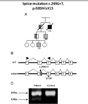
Alternatively spliced AIP transcript in the presence of the 249G > T mutation. A: Family tree (Family 28, Supp. Table S1) showing patients with gigantism (filled square), prolactinoma (striped squares), and carrier subjects (half-filled symbols) with age of onset shown. B: Schematic representation of splicing in the WTand mutant gene showing the location of the mutation (arrow) and the 32 bp lost from the end of exon 2 (shaded area) followed by a novel stop-codon after 15 novel codons (marked with *). Numbers in boxes represent exon numbers. Primers used are shown by arrowheads. C: RT-PCR using a patient and control cDNA. Patient cDNA shows an extra band (425 bp) that corresponds to the alternatively spliced AIP transcript with the upper band showing the WT transcript (457 bp). The identity of these PCR products were confirmed by sequencing.
Figure 3
Figure 3
Decreased AIP mRNA expression in the presence of the 807C > T. A: Family tree showing two patients with acromegaly and an asymptomatic carrier with age of onset of disease (Family XVI, Supp. Table S2). The location of the mutation is shown by an arrow; numbers in boxes represent exon numbers. B: Conventional RT-PCR with AIP primers on blood-derived cDNA obtained from three control individuals (C1–3) and two subjects (M1 and 2) carrying the 807C > T AIP mutation. Patients carrying the 807C > T showed decreased AIP expression. C: Real-time PCR using an AIP primer and probe set to compare the AIP levels between control individuals and patients; C (control). D: Wild-type (WT) and mutant minigene constructs. E: Conventional RT-PCR using vector-specific primers for the minigene constructs showing decreased mutated minigene expression (807T) and intermediate level of expression for the coexpression of WTand mutant minigenes (807CT). F: Real-time PCR showing an increased AIP expression in the WT minigene construct compared to mutant and coexpression of WTand mutant minigenes. EV, empty vector.
Figure 4
Figure 4
Single amino acid substitutions in AIP. A: A yeast two-hybrid quantitative β-galactosidase assay was used to assess the interaction of AIP with PDE4A5. It shows more than fivefold difference (activity 0–20% of wild-type [WT]) from WT AIP for missense mutations K103R, C238Y, K241E, and R271Was well as for the positive control truncation mutation Q164X. The R16H, V49M, I257V, A299V, and the R304Q variants show no difference or activity 33–100% of wild type (less than threefold difference) from WT AIP (mean ± SD). β-Galactosidase activity was measured as described by Guarente (1983). Each mutation was tested in at least two different yeast clones, with identical results (n = 3 for each clone). B: Hypothetical structure of AIP based on the structure of FKBP51 showing the three tetratricopeptide (TPR) domains with three pair of antiparallel α-helices and the final extended α-helix, α-7 (courtesy of Prof. David Barford, London, UK). Sequence comparison of human AIP and FKBP51 is shown in Supp. Figure S1. Amino acids with reported missense variants are highlighted. C: The three TPR domains, each consisting of an A and B helix, are shown of several Hsp90 binding proteins including AIP (table modified from Hidalgo-de-Quintana et al. [2008]). Numbers in diamond shapes show TPR motif position numbers. Amino acids marked with blue bold letters are important for the packaging and stability of the α-helices. Amino acids at position 8 and 20 were shown to be important in helix A and B packaging, while position 27 helps packaging of helix B with helix A of the same TPR domain and helix A of the following TPR domain. Underlined amino acids are predicted to be involved in the peptide binding pocket of FKBP51. Full-length variants of AIP affecting the TPR domains are shown on the figure (see review [Tahir et al., 2010]). Amino acids circled with red in the AIP sequence show missense variants described (C238Y, K241E, I257V, R271W). Amino acids marked with orange bold italics have been shown to be replaced by stop codon in FIPA patients (K201X, Q239X, K241X, Y268X). The amino acid marked by a red arrow is followed by an in-frame insertion (p.F269_H275dup) in a FIPA family. In-frame deletion variants p.Y238del is shown by strikethrough and complex missense and in-frame deletion mutation p.[E293G; L294_A297del] is shown by black circle and strikethrough.

References

    1. Beckers A, Daly AF. The clinical, pathological, and genetic features of familial isolated pituitary adenomas. Eur J Endocrinol. 2007;157:371–382. - PubMed
    1. Beckers A, Vanbellinghen JF, Boikos S, Martari M, Verma S, Daly AF, Raygada M, Keil M, Papademetriou J, Drori-Herishanu L, Horvath A, Nesterova M, Tichomirowa MA, Bours V, Marx S, Agarwal SK, Salvatori R, Stratakis CA. 2008. Germline AIP, MEN1, PRKAR1A, CDKN1B (p27Kip1) and CDKN2C (p18INK4c) gene mutations in a large cohort of pediatric patients with pituitary adenomas occurring in isolation or with associated syndromic features. Proc of the 90th Annual Meet of the Endocrine Soc; OR38-1.
    1. Bertherat J, Chanson P, Montminy M. The cyclic adenosine 3′,5′-monophosphate-responsive factor CREB is constitutively activated in human somatotroph adenomas. Mol Endocrinol. 1995;9:777–783. - PubMed
    1. Bolger GB, Peden AH, Steele MR, MacKenzie C, McEwan DG, Wallace DA, Huston E, Baillie GS, Houslay MD. Attenuation of the activity of the cAMP-specific phosphodiesterase PDE > A5 by interaction with the immunophilin XAP2. J Biol Chem. 2003;278:33351–33363. - PubMed
    1. Buchbinder S, Bierhaus A, Zorn M, Nawroth PP, Humpert P, Schilling T. Aryl hydrocarbon receptor interacting protein gene (AIP) mutations are rare in patients with hormone secreting or non-secreting pituitary adenomas. Exp Clin Endorcrinol Diabetes. 2008;116:625–628. - PubMed

Publication types

MeSH terms

Substances