. 2010 Jul 15;518(14):2729-43.
doi: 10.1002/cne.22361.

Cellular and subcellular localization of estrogen and progestin receptor immunoreactivities in the mouse hippocampus

Affiliations

Cellular and subcellular localization of estrogen and progestin receptor immunoreactivities in the mouse hippocampus

Katherine L Mitterling et al. J Comp Neurol. .

Abstract

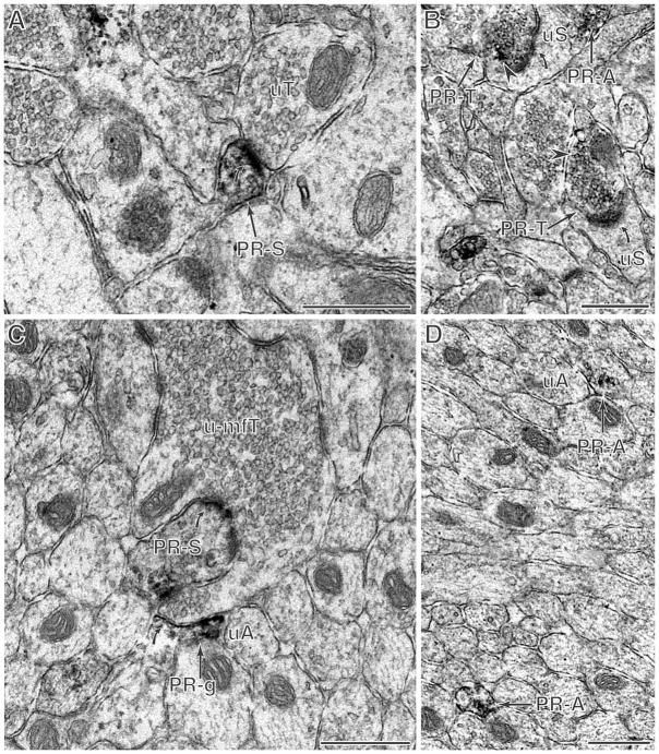
Estrogen receptor-alpha (ERalpha), estrogen receptor-beta (ERbeta), and progestin receptor (PR) immunoreactivities are localized to extranuclear sites in the rat hippocampal formation. Because rats and mice respond differently to estradiol treatment at a cellular level, the present study examined the distribution of ovarian hormone receptors in the dorsal hippocampal formation of mice. For this, antibodies to ERalpha, ERbeta, and PR were localized by light and electron immunomicroscopy in male and female mice across the estrous cycle. Light microscopic examination of the mouse hippocampal formation showed sparse nuclear ERalpha and PR immunoreactivity (-ir) most prominently in the CA1 region and diffuse ERbeta-ir primarily in the CA1 pyramidal cell layer as well as in a few interneurons. Ultrastructural analysis additionally revealed discrete extranuclear ERalpha-, ERbeta-, and PR-ir in neuronal and glial profiles throughout the hippocampal formation. Although extranuclear profiles were detected in all animal groups examined, the amount and types of profiles varied with sex and estrous cycle phase. ERalpha-ir was highest in diestrus females, particularly in dendritic spines, axons, and glia. Similarly, ERbeta-ir was highest in estrus and diestrus females, mainly in dendritic spines and glia. Conversely, PR-ir was highest during proestrus, mostly in axons. Except for very low levels of extranuclear ERbeta-ir in mossy fiber terminals in mice, the labeling patterns in the mice for all three antibodies were similar to the ultrastructural labeling found previously in rats, suggesting that regulation of these receptors is well conserved across the two species.


Figures

Figure 1
Figure 1
By light microscopy, ERα-, ERβ–, and PR-immunoreactivities are detected in the dorsal mouse hippocampal formation. A. Schematic diagram showing the hippocampal regions sampled for light and electron microscopy [adapted from (Hof et al., 2000)]. B–D. Examples of light microscopic localization of ERα-, ERβ–, and PR-immunoreactivities in the mouse CA1 region are shown. When the tissue is permeabilized with triton, sparse nuclear labeling for ERα is detected in stratum oriens (SO, arrows, B) and for PR in stratum radiatum (SR, arrows, D). Diffuse immunoreactivity for ERβ is detected in the pyramidal cell layer (pcl) and in a few interneurons (arrows, C). Bar 50 μm
Figure 2
Figure 2
ERα immunoreactivity is found in select dendrites, axons, terminals, and glia in males and females at all stages of the estrous cycle. A. ERα labeling is seen in a dendritic shaft at the base of a spine (ERa-S; arrowhead). (male mouse, DG mml) B. An ERα-labeled dendritic spine (ERa-S) receives a synapse (curved arrow) from an unlabeled terminal (uT). An unlabeled dendritic spine (uS) is shown for comparison. (diestrus, CA1 sr) C. An ERα-labeled terminal (ERa-T; arrowhead pointing at patch of peroxidase labeling) forms a synapse (curved arrow) with an unlabeled dendritic spine (uS). An unlabeled terminal (uT) and unlabeled dendritic shaft (uD) are nearby. (proestrus, CA3 so) D. In this bundle of axons, ERα labeling is found in a discrete patch within an unmyelinated axon (ERa-A) cut longitudinally. An unlabeled axon (uA) is also shown for comparison. (male, CA3 slu) E. ERα labeling is found in an unmyelinated pre-terminal axon leading to a terminal (T). (male, CA3 sr) F. ERα labeling was also found in a myelinated axon (ERa-mA; arrowhead). (Di, CA3 so) G. An ERα-labeled glial processes (ERa-g) apposes an unlabeled dendritic spine (uS) and an unlabeled terminal (uT). (pro, CA1 sr) Bar 500nm.
Figure 3
Figure 3
ERβ immunoreactivity is found in select dendrites, axons, terminals, and glia in males and females at all stages of the estrous cycle. A. ERβ labeling is in discrete patches (examples, arrowheads) throughout the cytoplasm of a perikaryon. The higher magnification (inset) shows a dense patch of ERβ–labeling associated at an endomembrane near the mitochondrion (bar 250 nm). (CA1 sr) B. An ERβ-labeled spine (ERb-S) arising from a dendritic shaft lacking labeling (D) is contacted (curved arrow) by an unlabeled terminal (uT). (CA1 so) C. ERβ labeling is found in a dendrite (ERb-D), both in the cytoplasm (black arrowhead) and on a mitochondrion (white arrowhead). (CA1 sr) D. ERβ labeling is found in a glia process (ERb-g) that conforms to the boundaries of the neuropil. (CA1 sr) E. An ERβ-labeled terminal (ERb-T; arrowhead points to cluster of peroxidase labeling) forms a synapse (curved arrow) with an unlabeled dendritic spine (uS). (CA1 sr) F. ERβ-labeling is found in an astrocytic profile abutting the basement membrane (bm) of a blood vessel (bv). (CA1 sr near) Bar A–C and F 500nm; D and F 250nm
Figure 4
Figure 4
PR immunoreactivity is found in select dendrites, axons, terminals, and glia primarily in proestrus mice. A. A PR-labeled dendritic spine (PR-S) receives a synapse (curved arrow) from an unlabeled terminal (uT). (proestrus, DG sgz) B. Two PR-labeled terminals (PR-T; arrowhead) synapse (curved arrows) on unlabeled dendritic spines (uS). A PR-labeled unmyelinated axon (PR-A) apposes to one of the unlabeled dendritic spines. (Pro, CA1 sr dist) C. A PR-labeled spine (PR-S) is contacted (curved arrows) by an unlabeled mossy fiber terminal (u-mfT). An unlabeled axon (uA) apposes a PR-labeled glial process (PR-g). (pro, dg hil) D. Two PR-labeled unmyelinated axons (PR-A) are shown in a field of unlabeled unmyelinated axons. An unmyelinated axon (uA) is marked for comparison. (pro, dg hil) Bar 500nm
Figure 5
Figure 5
Summary diagram showing the levels of ERα-, ERβ, and PR-ir in male mice and across the estrous cycle in female mice in comparison to the known fluctuations of circulating estradiol and progesterone.

Similar articles

Cited by

References

    1. Akama KT, McEwen BS. Estrogen stimulates PSD-95 rapid protein synthesis via the Akt/PKB pathway. J Neurosci. 2003;23:2333–2339. - PMC - PubMed
    1. Alves SE, McEwen BS, Hayashi S, Korach KS, Pfaff DW, Ogawa S. Estrogen-regulated progestin receptors are found in the midbrain raphe but not hippocampus of estrogen receptor alpha (ERα) gene-disrupted mice. J Comp Neurol. 2000;427:185–195. - PubMed
    1. Alves SE, Weiland NG, Hayashi S, McEwen BS. Immunocytochemical localization of nuclear estrogen receptors and progestin receptors within the rat dorsal raphe nucleus. J Comp Neurol. 1998;391:322–334. - PubMed
    1. Arias C, Zepeda A, Hernandez-Ortega K, Leal-Galicia P, Lojero C, Camacho-Arroyo I. Sex and estrous cycle-dependent differences in glial fibrillary acidic protein immunoreactivity in the adult rat hippocampus. Horm Behav. 2009;55:257–263. - PubMed
    1. Becker JB, Hu M. Sex differences in drug abuse. Front Neuroendocrinol. 2008;29:36–47. - PMC - PubMed

Publication types

MeSH terms

Substances