. 2010 Jul 15;518(14):2818-40.
doi: 10.1002/cne.22366.

Identification of cerebellin2 in chick and its preferential expression by subsets of developing sensory neurons and their targets in the dorsal horn

Affiliations

Identification of cerebellin2 in chick and its preferential expression by subsets of developing sensory neurons and their targets in the dorsal horn

Mao Yang et al. J Comp Neurol. .

Abstract

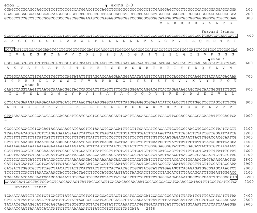
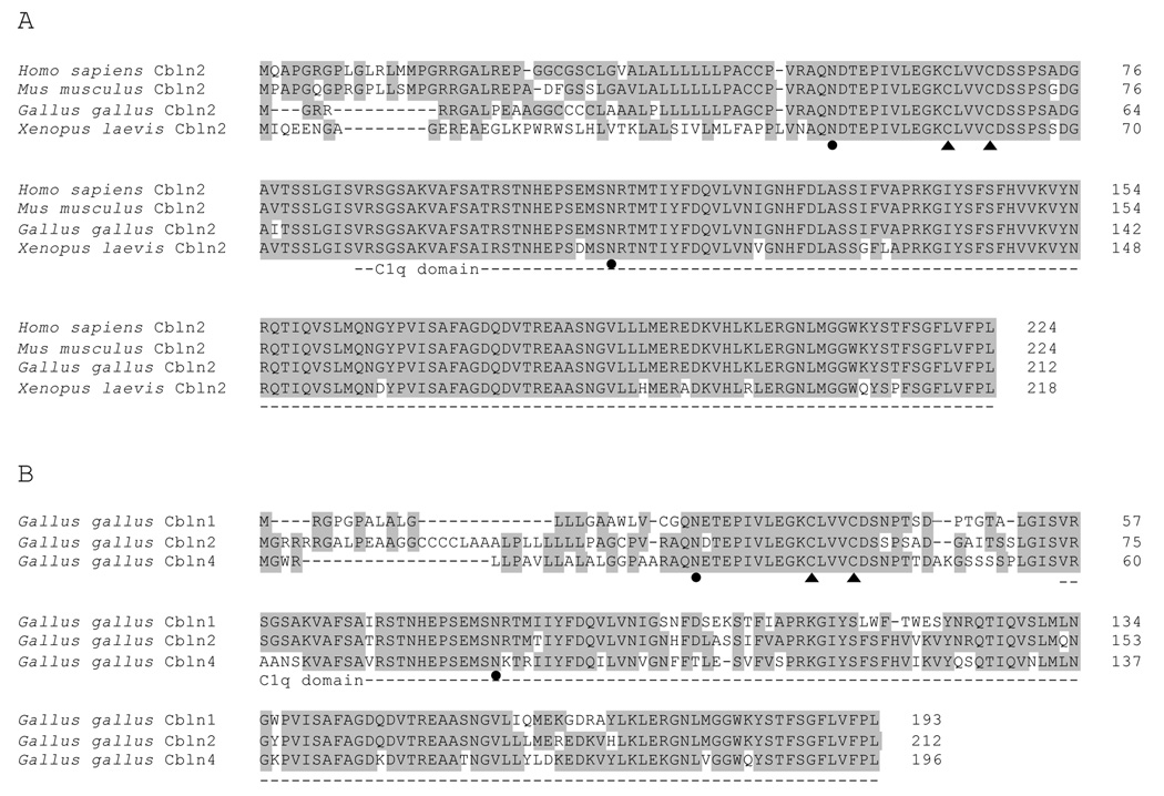
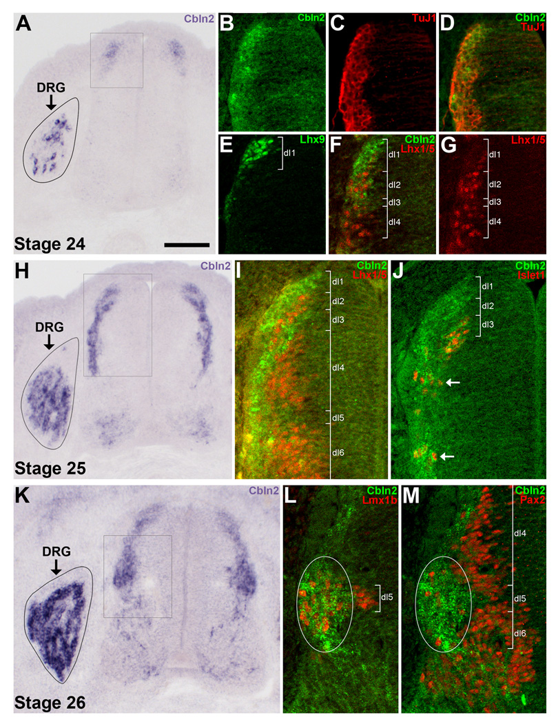
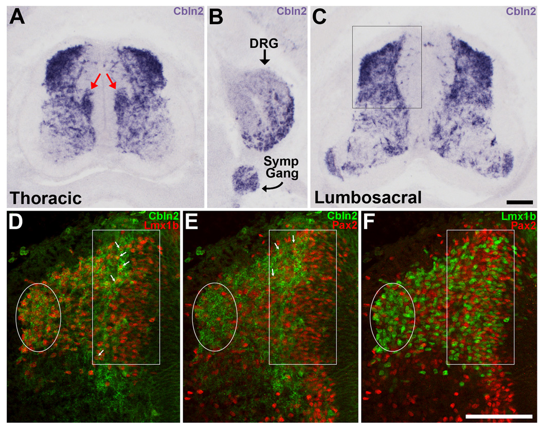
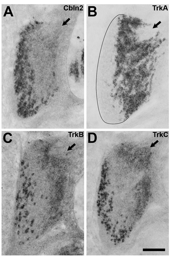
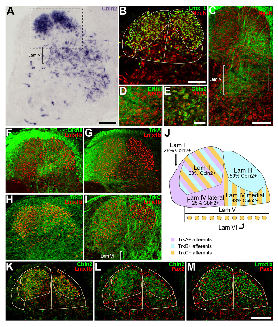
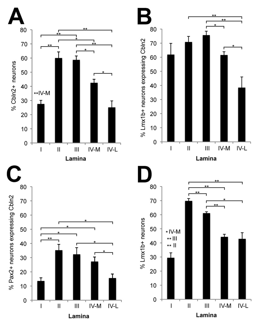
The cerebellins are a family of four secreted proteins, two of which, Cbln1 and Cbln3, play an important role in the formation and maintenance of parallel fiber-Purkinje cell synapses. We have identified the chicken homologue of Cbln2 and, through the use of in situ hybridization, shown that it is expressed by specific subsets of neurons in the dorsal root ganglia (DRGs) and spinal cord starting shortly after those neurons are generated. In the developing spinal cord, Cbln2 is highly expressed by dI1, dI3, dI5, and dILB dorsal interneurons and to a lesser extent by dI2, dI4, dI6, and dILA dorsal interneurons, but not by ventral (v0-v3) interneurons. After the spinal cord has matured and neurons have migrated to their final destinations, Cbln2 is abundant in the dorsal horn. In the DRGs, Cbln2 is expressed by TrkB+ and TrkC+ sensory neurons, but not by TrkA+ sensory neurons. Interestingly, regions of the spinal cord where TrkB+ and TrkC+ afferents terminate (i.e., laminae II, III, IV, and VI) exhibit the highest levels of Cbln2 expression. Cbln2 is also expressed by preganglionic sympathetic neurons and their targets in the sympathetic chain ganglia. Thus, the results show that Cbln2 is frequently expressed by synaptically connected neuronal populations. This, in turn, raises the possibility that if Cbln2, like Cbln1, plays a role in the formation and maintenance of synapses, it may somehow mediate bi-directional communication between discrete populations of neurons and their appropriate neuronal targets.


Figures

Figure 1
Figure 1. Cbln2 from Gallus gallus
The open reading frame that encodes chicken Cbln2 is underlined, and the predicted amino acid sequence is shown. The forward and reverse primers used for PCR are indicated. The inverted triangles mark the intron-exon splicing junctions predicted by comparing the mRNA sequence and the genomic sequence. Note that the junction between exon 2 and exon 3 has not been determined because the genomic sequence in this region is incomplete.
Figure 2
Figure 2. Cbln2 from Gallus gallus is highly homologous to Cbln2 from other species and to Cbln1 and Cbln4 from Gallus gallus
A) Cbln2 from human (Genbank accession number NP_872317), mouse (Genbank accession number NP_766221), chicken (translated from Genbank accession number GU189513), and frog (Genbank accession number NP_001080811) are highly conserved, especially in the C1q domain (dotted line). B) Cbln2 from Gallus gallus shares homology with other cerebellin family members, Cbln1 (translated from Genbank accession number XM_001233212, modified as described in Materials and Methods) and Cbln4 (Genbank accession number NP_001072955). Cbln3 is believed to be absent in Gallus gallus. Conserved amino acid residues are shown in gray. The conserved cysteine residues for oligomerization are indicated by filled triangles. The conserved asparagines residues for N-glycosylation are indicated by filled circles.
Figure 3
Figure 3. In situ hybridization for Cbln2 in the spinal cord and DRGs at St. 24–26
A–G) Cbln2 expression at St. 24. A) Cbln2 is expressed by several neurons in the incipient DRGs and at the far dorsal edge of the spinal cord. B–G) Only the dorsal part of the left side of the spinal cord (indicated by the box in panel A) is shown. B–D) Cbln2+ cells at the far dorsal edge of the spinal cord are TuJ1+. E) Lhx9+ dI1 neurons are situated at the far dorsal edge of the spinal cord. F) Cbln2 is predominantly expressed in the region just dorsal to Lhx1/5+ dI2 neurons, by neurons in the same position as the Lhx9+ dI1 neurons shown in panel E. G) The same section as in panel F, showing immunofluorescent staining for Lhx1/5 alone to visualize dI2 and dI4 neurons. H–J) Cbln2 expression at St. 25. H) Cbln2 is expressed by neurons spread along the lateral border of the dorsal half of the spinal cord, by some neurons in the ventral cord, and by an increasing number of DRG neurons. I–J) Only part of the left side of the spinal cord (indicated by the box in panel H) is shown. I) Cbln2 is expressed by a few of the most laterally-situated Lhx1/5+ dI2, dI4 and dI6 neurons. There is a gap in Cbln2 labeling in the dI2 region. J) Cbln2 is expressed by most Islet1+ dI3 neurons, a few of which have already migrated ventrally (arrows). K–M) Cbln2 expression at St. 26. K) Cbln2 is expressed by numerous DRG neurons. In the spinal cord, many dI1 and dI3 neurons (located above the boxed region) express Cbln2, as they do at St. 25, but Cbln2 expression has now expanded to also include a large cluster of neurons (situated within the boxed region). L–M) Only part of the left side of the spinal cord (indicated by the box in panel K) is shown. Cbln2 is expressed by the several Lmx1b+ dI5 neurons and the few Pax2+ dI4/dI6 neurons that are situated within this lateral cluster (white ovals). For all images, dorsal is up. For A, H, and K the DRG on the left side only is shown due to space considerations. For B–D, F–G, I–J, and L–M, in situ hybridization for Cbln2 was followed by processing for immunofluorescence. Note that Cbln2 mRNA is localized to the cytoplasm whereas the transcription factors are expressed in the nucleus, and so double-labeled neurons do not necessarily appear yellow. Magenta-green version available as Supporting Figure 3. Scale bar in A is 100µm for panels A, H, and K and 50µm for panels B–G, I–J, L–M.
Figure 4
Figure 4. In situ hybridization for Cbln2 in the spinal cord at St. 30
A) At thoracic levels, Cbln2 is heavily expressed in the dorsal part of the spinal cord and by sympathetic preganglionic neurons in the column of Terni (red arrows). B) In the same section as in (A), Cbln2 is expressed in the DRGs and in the sympathetic chain ganglia. Medial is to the left. C) In lumbosacral segments, Cbln2 is heavily expressed in the dorsal half of the spinal cord and by some motoneurons. D–F) The dorsal part of the left side of the spinal cord (indicated by the box in panel C) is shown. The same section is shown in all three panels. The lateral cluster of dI4–6 neurons has migrated dorsally (enclosed in white oval) and is now situated at the dorsolateral edge of spinal cord. Numerous dILA and dILB neurons are now present and located medially (white rectangle). D, E) Some of the more lateral Lmx1b+ dILB and Pax2+ dILA neurons express Cbln2 (arrows). F) Lmx1b+ dILB and Pax2+ dILA are intermixed in a salt-and-pepper-like manner. Magenta-green version available as Supporting Figure 4. Scale bar, 100µm in C is for panels A, B, and C; in F is for panels D, E and F.
Figure 5
Figure 5. Cbln2 is expressed by TrkB+ and TrkC+ DRG neurons, but not by TrkA+ DRG neurons. In situ hybridization on sections from St. 30 embryos
A) Cbln2 is expressed primarily by large neurons in the ventral and lateral parts of the DRG. B) TrkA is expressed by small neurons in the dorsomedial part of the DRG. C) TrkB is expressed by large neurons situated amongst and slightly medial to the TrkC+ neurons. D) TrkC is expressed by large neurons situated at the lateral edge of the DRG. Low levels of TrkB and TrkC expression in small neurons in the dorsomedial region were not considered in our analysis. Some of the small neurons in the dorsomedial region expressing low levels of TrkB or TrkC express high levels of TrkA and were therefore considered to be TrkA+ in our analysis. The dorsomedial pole of each DRG is marked with an arrow. Dorsal is toward the top, lateral to the left. Scale bar, 100µm.
Figure 6
Figure 6. At St. 37, Cbln2 is expressed in regions of the dorsal and intermediate spinal cord where TrkB+ and TrkC+ axons arborize
A) Cbln2 is most heavily expressed in the dorsal horn and in lamina VI. B) Laminae I–IV can be distinguished by immunofluorescent staining for Lmx1b and NeuN. See text for description and panel J for lamina identification. The area shown is equivalent to that indicated by the box in panel A. C) Sensory axons arborize most extensively in lamina IV and lamina VI. The dorsal roots (DR) were filled with Alexa 488-dextran. D) Higher magnification view of the box outlined in panel C. Sensory axons arborize amidst the large neurons in lamina VI. E) Cbln2 is expressed by large neurons in lamina VI. The area shown is from a different embryo but is similar to that shown in panel D. F–I) Sensory axon projections into the dorsal spinal cord. Sections were also labeled for Lmx1b to aid in distinguishing laminae. F) Sensory axons filled with Alexa 488-dextran arborize in laminae I, II, III, and IV. G) TrkA+ afferents terminate in laminae I and II, and the lateral part of lamina IV. The same section as in panel F is shown. H) TrkB+ afferents arborize primarily in lamina III and the medial part of lamina IV, and to a lesser extent, in the dorsolateral part of lamina II. I) TrkC+ afferents arborize in lamina II, the medial part of lamina IV, and in lamina VI. TrkC+ axons cross lamina III on their way to lamina IV, but it is not clear if any of them terminate in lamina III. In some embryos, light labeling for TrkC and for TrkB could be seen in lamina I. J) Schematic overview of laminae I–VI. Colors indicate where TrkA+, TrkB+, and TrkC+ axons arborize. The percentage of neurons in each lamina that expresses Cbln2 is indicated. K–L) Cbln2 is most abundant in lamina II and lamina III and expressed at lower levels in lamina I and lamina IV. K) Many Lmx1+ neurons express Cbln2. L) Some, albeit fewer, Pax2+ neurons express Cbln2. M) Lmx1+ and Pax2+ neurons are intermixed in laminae I–IV. Magenta-green version available as Supporting Figure 5. Scale bars, 100µm in A, C, M; 50µm in B and E. Scalebar in M also applies to panels F–I, K–L. Scale bar in E also applies to panel D.
Figure 7
Figure 7. Cbln2 is mainly expressed in lamina II and lamina III of the dorsal horn and more frequently by the excitatory Lmx1b+ neurons than by the Pax2+ inhibitory neurons in dorsal horn
Cbln2+ neurons in the dorsal horn were counted in six sections that were processed first for in situ hybridization for Cbln2, and then for immunofluorescence to visualize Lmx1b+ and Pax2+ neurons. A) The percentage of neurons that are Cbln2+ is greatest in lamina II and lamina III, less in lamina IV medial, and lowest in lamina I and lamina IV lateral. B) The percentage of Lmx1b+ neurons that are Cbln2+ is lowest in lamina IV lateral. C) The percentage of Pax2+ neurons that are Cbln2+ is lowest in lamina I and in lamina IV lateral. D) The percentage of neurons that are Lmx1b+ is lowest in lamina I and is lower in lamina IV medial and lamina IV lateral than in lamina II or lamina III. Values shown are mean ± standard error of the mean. Statistically significant differences between laminae were determined using two-tailed Mann-Whitney U tests and are indicated either by brackets indicating each comparison or, to minimize excessive crowding, above the bars showing the lamina I values. Probability is indicated by the number of asterisks (*P ≤ 0.05; ***P ≤ 0.005). The significance levels (p values) and the statistic (U) values for these comparison are provided in Supporting Table 1.

Similar articles

Cited by

References

    1. Airaksinen MS, Koltzenburg M, Lewin GR, Masu Y, Helbig C, Wolf E, Brem G, Toyka KV, Thoenen H, Meyer M. Specific subtypes of cutaneous mechanoreceptors require Neurotrophin-3 following peripheral target innervation. Neuron. 1996;16:287–295. - PubMed
    1. Bao D, Pang Z, Morgan JI. The structure and proteolytic processing of Cbln1 complexes. J Neurochem. 2005;95:618–629. - PubMed
    1. Bao D, Pang Z, Morgan MA, Parris J, Rong Y, Li L, Morgan JI. Cbln1 is essential for interaction-dependent secretion of Cbln3. Mol Cell Biol. 2006;26:9327–9337. - PMC - PubMed
    1. Barrow SL, Constable JR, Clark E, El-Sabeawy F, McAllister AK, Washbourne P. Neuroligin1: a cell adhesion molecule that recruits PSD-95 and NMDA receptors by distinct mechanisms during synaptogenesis. Neural Dev. 2009;4:17. - PMC - PubMed
    1. Bermingham NA, Hassan BA, Wang VY, Fernandez M, Banfi S, Bellen HJ, Fritzsch B, Zoghbi HY. Proprioceptor pathway development is dependent on MATH1. Neuron. 2001;30:411–422. - PubMed

Publication types

MeSH terms