. 2010 Jul;19(7):1366-75.
doi: 10.1002/pro.415.

How sequence directs bending in tropomyosin and other two-stranded alpha-helical coiled coils

Affiliations

How sequence directs bending in tropomyosin and other two-stranded alpha-helical coiled coils

Jerry H Brown. Protein Sci. 2010 Jul.

Abstract

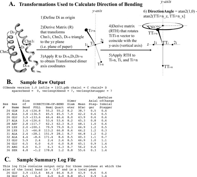
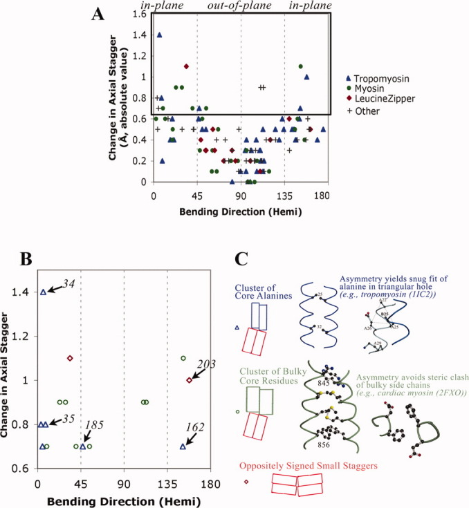
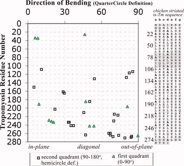
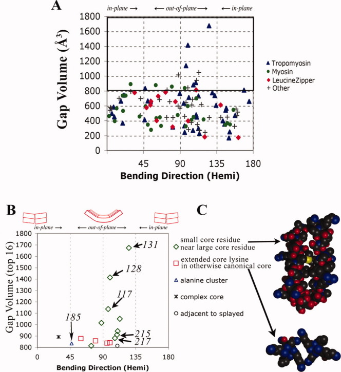
A quantitative analysis of the direction of bending of two-stranded alpha-helical coiled coils in crystal structures has been carried out to help determine how the amino acid sequence of the coiled coil influences its shape and function. Change in the axial staggering of the coiled coil, occurring at the boundaries of either clusters of core alanines in tropomyosin or of clusters of core bulky residues in the myosin rod, causes bending within the plane of the local dimer. The results also reveal that large gaps in the core of the coiled coil, which are seen for small core residues near large core residues or for unbranched core residues near canonical branched core residues, are correlated with bending out of the local dimeric plane. Comparison of tropomyosin structures determined in independent crystal environments provides further evidence for the concept that sequence directs the bending of the coiled coil, but that crystal environment is at least as important as sequence for determining the magnitude of bending. Tropomyosin thus appears to consist of more directionally restrained hinge-like joints rather than directionally variable universal joints, which helps account for and predicts the geometric and dynamic nature of its binding to F-actin.


Figures

Figure 1
Figure 1
Direction of bending calculation and output files of CCBENDS. A: The indicated transformations facilitate calculation of the fully signed bending direction angle of a parallel dimeric alpha-helical coiled coil axis using the atan2 function. Value of 0° indicates bending of the axis within the local plane towards chain 1, ±180° indicate in-plane bending towards chain 2, ±90° indicate out-of-plane bending, and ±45° or ±135° indicate diagonal bending (see also Table SI in Supporting Information). The example schematic shown in this figure displays a direction of bending of c. +70°, nearly out of the plane of the local dimer. (Note that the chain axis at each residue is calculated using the coordinates of the nearest five alpha-carbons, and the dimer axis is the simple average of the chain axes.) B: Sample raw output file includes fully signed and symmetry-reduced (“hemi” or “quart”) local bend direction angles, as well as other coiled-coil parameters for each residue (See Methods for explanations). C: Corresponding summary file extracts from the raw output file those residues at which the size of the bend is both >3.0° and is a local peak, that is, greater than that of the prior and subsequent residue. [In this example, the fully signed measurement shows that the two local bends, which bracket an axially staggered stretch, are oppositely signed (i.e., closer to 180° apart than 0° apart), which is a signature of this (near) in-plane bending design. Such pairs of nearly oppositely signed near-in-plane local bending angles also surround three other axially staggered alanine clusters in tropomyosin as well as axially staggered clusters of bulky core residues in myosin. See also Fig. 3.]
Figure 3
Figure 3
Large changes in axial staggering of the α-helices promote in-plane bending. A: Scatter plot of results from summary log files of all relatively high resolution crystal structures of parallel homodimeric coiled coil according to SCOP as of January 15, 2009. In addition to tropomyosin (see Fig. 2), coiled coils are from myosin (pdb i.d. 1nkn; 3bas; 3bat; 2fxm; 2fxo), leucine zipper domain (2zta, 1zik, 2ahp, 1zil, 1pyI, 1zme, 1hwt, 2hap, 1qp9, 1gd2, 1gu4, 1gtw, 1h8A, 1h88, 1hjb, 1io4, 1nwq, 2c9l, 2c9n), and other proteins (1dkg, 1d7m, 1deb, 1joc, 1no4, 1noh, 1uix, 1s1c, 1x79, 1tu3, 1T6F, 1uii, 2gzd, 2gzh, 2d7c, 2hv8, and 2ocy) (see also Table SIII). The hemicircle-reduced direction of bending is shown on the x-axis. At each position along the sequence, i, the change in axial stagger per heptad = [axial stagger at position (i+3) + stagger at position (i+4) − stagger at (i−3) − stagger at (i−4)]/2.0, and the absolute value is printed out on the y-axis. B: The 16 bends with greatest change in axial staggering. Italicized numbers are tropomyosin residues. C: Two major designs that promote axial staggering. See also text.
Figure 2
Figure 2
Direction of bending is relatively conserved at most locations of tropomyosin. Scatter plot of all 39 local bends [from summary log files as in Fig. 1(C)] found in crystal structures of fragments of vertebrate striated alpha-tropomyosin (pdb i.d. 1ic2, 2b9c, 1kql, 2d3e, 2efr, 2efs, 2z5h, 2z5i: see also Table SII in Supporting Information), with the x-axis showing the quartercircle-reduced direction of bending. [Only those residues more than seven residues from a non-tropomyosin or non-coiled-coil region are included. The program was run separately on each of the crystallographically independent copies of the coiled coil, except for chains CD of 2d3e and chains EF and GH of 2z5I, which have nearly identical (<0.1 RMS different) conformations to those included.] Twenty-three of the bends are from eight locations in which bending occurs in more than one crystal environment (see text). Sequence (see right panel) appears to be the more important factor controlling the direction of bends of the tropomyosin coiled coil, whereas crystal environment play at least as important a role for determining the magnitude of the bend (see text and Fig. S1 in Supporting Information). Shaded sequences have not been examined by CCBENDS because either there is no crystal structure available or because the position is within 10 residues of a terminus of the structure. [Color figure can be viewed in the online issue, which is available at www.interscience.wiley.com.]
Figure 4
Figure 4
Large gaps at the interhelical interface promote out-of-plane bending. A: Same database of structures is used as in Figure 3. For each bend at residue i listed in the summary log file, the coordinates of residues i−7 to i+7 from each chain were extracted using PDBCUR from the CCP4 suite, and the gap volume was obtained using the Protein–Protein Interface Comparison server. Over all 117 bends, the median gap volume is 567 Å. B: The 16 bends with the largest gap volumes. Tropomyosin residues 128, 131, and 117 are from pdb i.d. 2b9c, 185, and 215 are from 2d3e, and 217 is from 2efr. C: Two types of gap-containing structures in which the main chains are propped apart at the locations of small or medially unbranched side chains. See also text.

References

    1. O'Shea EK, Klemm JD, Kim PS, Alber T. X-ray structure of the GCN4 leucine zipper, a two-stranded, parallel coiled coil. Science. 1991;254:539–544. - PubMed
    1. Burkhard P, Stetefeld J, Strelkov SV. Coiled coils: a highly versatile protein folding motif. Trends Cell Biol. 2001;11:82–88. - PubMed
    1. Brown JH, Cohen C. Regulation of muscle contraction by tropomyosin and troponhow structure illuminates function. In: Squire JM, Parry DA, editors. Fibrous proteins and related structures. Elsevier/Academic press; 2005.
    1. Crick FHC. The packing of alpha-helices: simple coiled coils. Acta Crystallogr. 1953;6:689–697.
    1. Cohen C, Parry DAD. Alpha-helical coiled coils and bundles: how to design an alpha-helical protein. Protein Struct Funct Genet. 1990;7:1–15. - PubMed

Publication types

LinkOut - more resources