. 2010 Jun;19(6):1222-34.
doi: 10.1002/pro.401.

A computational study of RNA binding and specificity in the tandem zinc finger domain of TIS11d

Affiliations

A computational study of RNA binding and specificity in the tandem zinc finger domain of TIS11d

Brittany R Morgan et al. Protein Sci. 2010 Jun.

Abstract

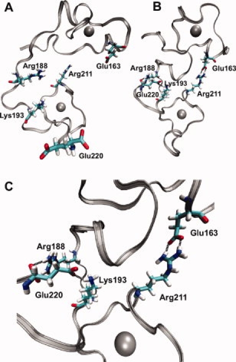
TIS11d is a member of the CCCH-type family of tandem zinc finger (TZF) proteins; the TZF domain of TIS11d (residues 151-220) is sufficient to bind and destabilize its target mRNAs with high specificity. In this study, the TZF domain of TIS11d is simulated in an aqueous environment in both the free and RNA-bound states. Multiple nanosecond timescale molecular dynamics trajectories of TIS11d wild-type and E157R/E195K mutant with different RNA sequences were performed to investigate the molecular basis for RNA binding specificities of this TZF domain. A variety of measures of the protein structure, fluctuations, and dynamics were used to analyze the trajectories. The results of this study support the following conclusions: (1) the structure of the two fingers is maintained in the free state but a global reorientation occurs to yield a more compact structure; (2) mutation of the glutamate residues at positions 157 and 195 to arginine and lysine, respectively, affects the RNA recognition by this TIS11d mutant in agreement with the findings of Pagano et al. (J Biol Chem 2007; 282:8883-8894); and (3) we predict that the E157R/E195K mutant will present a more relaxed RNA binding specificity relative to wild-type TIS11d based on the more favorable nonsequence-specific Coulomb interaction of the two positively charged residues at positions 157 and 195 with the RNA backbone, which compensates for a partial loss of the stacking interaction of aromatic side chains with the RNA bases.


Figures

Figure 1
Figure 1
A: Schematic representation of the TZF domain of TIS11d; ZF1, ZF2, and the linking region are indicated with boxes and residue numbers. B: Sequence alignment of CCCH-type tandem zinc finger proteins. Alignment of human (h) TTP with hTIS11d/b and C. elegans (c) MEX-5, MEX-6, MEX-1, POS-1, and PIE-1. Conserved motifs (R(K)YTEL) are represented in blue. The three cysteines and the histidine involved in zinc binding are highlighted in red. Residues directly involved in RNA binding in the NMR structure of TIS11d are indicated with an asterisk: a black asterisk is used for residues which form a hydrogen bond with a base of the RNA sequence and a green asterisk identifies aromatic residues involved in base-stacking interactions with the RNA. C: NMR solution structure of the TIS11d-ARE complex (PDB accession code 1RGO). Pink: TIS11d with zinc-coordinating residues shown, gray spheres: zinc ions, purple: RNA.
Figure 2
Figure 2
Compaction of TIS11d structure. A: Snapshots colored by time. Left: TIS11d-ARE between 10 and 14 ns aligned on the protein backbone—red (10 ns), orange (10.4 ns), yellow (10.8 ns), green (11.6 ns), cyan (12 ns), blue (12.8 ns), indigo (13.6 ns), purple (14 ns), right: TIS11d between 24 and 28 ns aligned on the protein backbone—red (24 ns), orange (24.4 ns), yellow (24.8 ns), green (25.6 ns), cyan (26 ns), blue (26.8 ns), indigo (27.6 ns), purple (28 ns). B, C: Red: NMR structure of the TIS11d-ARE complex depicted without showing the RNA (PDB accession code 1RGO), blue: structures from MD trajectories aligned on NMR structure (see below), gray spheres: zinc ions. B: Snapshot of the TIS11d-ARE complex at 14 ns. C: Snapshot of TIS11d at 28 ns. From left to right: full TZF domain, ZF1, and ZF2. Full TZF domain is aligned on all backbone atoms (B) or the backbone atoms of ZF1 (C), ZF1 and ZF2 are aligned on their respective backbone atoms. D: Normalized histograms of the total protein solvent accessible surface area (SASA). SASA of the TIS11d-ARE complex is represented in black, when the RNA treated as solvent, and red when the RNA shielding is included. SASA calculated for two MD trajectories of TIS11d are represented in blue and green.
Figure 3
Figure 3
Difference of the mean order parameter by residue. Error bars are calculated from the standard deviation among trajectories. The boxes in (A) highlight the two zinc finger domains. A: formula imageformula image; B: formula imageformula image.
Figure 4
Figure 4
Hydrogen bonds (black dashed lines) between different finger domains of TIS11d. Silver ribbon: protein backbone, gray spheres: zinc ions, residues as labeled. A: The structure of TIS11d-ARE (RNA not shown) at 14 ns. B: Structure of TIS11d at 28 ns. C: Structure of TIS11d at 28 ns enlarged to show detail.
Figure 5
Figure 5
Adenine recognition pocket of ZF2. Red: protein side chains for residues 195–198, blue: protein backbone, gray: RNA phosphate group and sugar of nucleotide 3, green: RNA base ring of nucleotide 3. Hydrogen bonds are depicted with a black dashed line. (A) TIS11d-ARE, (B) TIS11d-U9, (C) E157R/E195K-ARE, and (D) E157R/E195K-U9.
Figure 6
Figure 6
Structures of E157R/E195K mutants and different RNA sequences (RNA not shown). The zinc ion of each finger is represented in gray. Proteins are shown with ZF2 on the left and ZF1 on the right. From left to right: full TZF domain, ZF1, and ZF2, respectively aligned on NMR of the TIS11d-ARE complex structure (in red) using the backbone atoms of ZF1, ZF1, and ZF2. Blue (A) TIS11d-U9, (B) E157R/E195K-U9, and (C) E157R/E195K-ARE.
Figure 7
Figure 7
Protein–RNA hydrogen bond probabilities. The mean and standard deviation of the sum of all protein–RNA hydrogen bond probabilities is represented for each RNA nucleotide. Red: TIS11d-ARE, blue: TIS11d-U9, green: E157R/E195K-U9, and purple: E157R/E195K-ARE.
Figure 8
Figure 8
RNA intercalation. Red: protein side chains of residues 170,176, 208, and 214, blue: protein backbone, silver spheres: zinc ions, gray: RNA backbone and sugars, and green: RNA base rings. ZF2 is enlarged on the right to show detail. (A) NMR structure of TIS11d-ARE and (B) E157R/E195K-U9. (C) Mean and standard deviation of the distance between the geometric centers of the intercalating aromatic rings within each intercalating base stack. Red: TIS11d-ARE, blue: TIS11d-U9, green: E157R/E195K-U9, and purple: E157R/E195K-ARE.
Figure 9
Figure 9
Normalized histograms of protein–RNA Coulomb interaction energy for the N-terminal tail (residues 151–158) and linking residues (residues 179–196) combined. (A) Gray: TIS11d-ARE, black: TIS11d-U9, (B) gray: E157R/E195K-ARE, and black: E157R/E195K-U9.

Similar articles

Cited by

References

    1. Ogilvie RL, Abelson M, Hau HH, Vlasova I, Blackshear PJ, Bohjanen PR. Tristetraprolin down-regulates IL-2 gene expression through AU-rich element-mediated mRNA decay. J Immunol. 2005;174:953–961. - PubMed
    1. Carballo E, Lai WS, Blackshear PJ. Feedback inhibition of macrophage tumor necrosis factor-alpha production by tristetraprolin. Science. 1998;281:1001–1005. - PubMed
    1. Carrick DM, Lai WS, Blackshear PJ. The tandem CCCH zinc finger protein tristetraprolin and its relevance to cytokine mRNA turnover and arthritis. Arthritis Res Ther. 2004;6:248–264. - PMC - PubMed
    1. Chen CY, Gherzi R, Ong SE, Chan EL, Raijmakers R, Pruijn GJ, Stoecklin G, Moroni C, Mann M, Karin M. AU binding proteins recruit the exosome to degrade ARE-containing mRNAs. Cell. 2001;107:451–464. - PubMed
    1. Lai WS, Carballo E, Strum JR, Kennington EA, Phillips RS, Blackshear PJ. Evidence that tristetraprolin binds to AU-rich elements and promotes the deadenylation and destabilization of tumor necrosis factor alpha mRNA. Mol Cell Biol. 1999;19:4311–4323. - PMC - PubMed

MeSH terms