. 2011 Jan;112(1):39-48.
doi: 10.1002/jcb.22710.

Identification of phospholipase C gamma1 as a protein tyrosine phosphatase mu substrate that regulates cell migration

Affiliations

Identification of phospholipase C gamma1 as a protein tyrosine phosphatase mu substrate that regulates cell migration

Polly J Phillips-Mason et al. J Cell Biochem. 2011 Jan.

Abstract

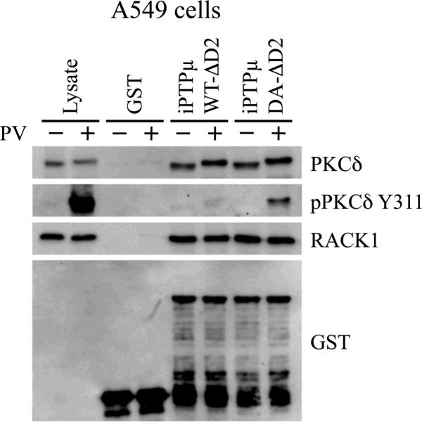
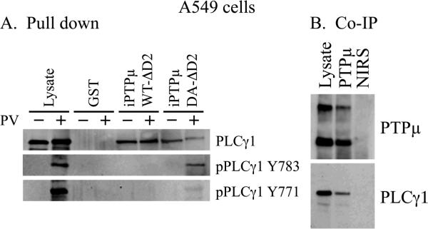
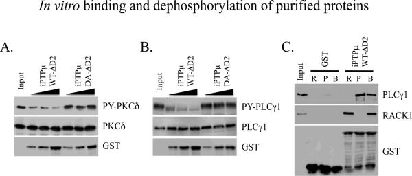
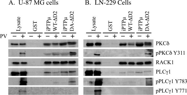
The receptor protein tyrosine phosphatase PTPµ has a cell-adhesion molecule-like extracellular segment and a catalytically active intracellular segment. This structure gives PTPµ the ability to transduce signals in response to cell-cell adhesion. Full-length PTPµ is down-regulated in glioma cells by proteolysis which is linked to increased migration of these cells in the brain. To gain insight into the substrates PTPµ may be dephosphorylating to suppress glioma cell migration, we used a substrate trapping method to identify PTPµ substrates in tumor cell lines. We identified both PKCδ and PLCγ1 as PTPµ substrates. As PLCγ1 activation is linked to increased invasion of cancer cells, we set out to determine whether PTPµ may be upstream of PLCγ1 in regulating glioma cell migration. We conducted brain slice assays using U87-MG human glioma cells in which PTPµ expression was reduced by shRNA to induce migration. Treatment of the same cells with PTPµ shRNA and a PLCγ1 inhibitor prevented migration of the cells within the brain slice. These data suggest that PLCγ1 is downstream of PTPµ and that dephosphorylation of PLCγ1 is likely to be a major pathway through which PTPµ suppresses glioma cell migration.


Figures

Figure 1
Figure 1
PKCδ is a substrate for PTPμ. A549 cells were treated with (+) or without (−) 100 μM pervanadate (PV) for 20 minutes. Cells were lysed and equal amounts of protein were incubated with iPTPμWT-ΔD2-GST, iPTPμDA-ΔD2-GST or GST immobilized on glutathione Sepharose. Associated proteins were resolved by SDS-PAGE (8% for PKCδ and 10% for RACK1) and detected by immunoblot. PKCδ and RACK1 were detected in all the PTPμ pull downs. Phospho-PKCδ Y311 was detected only in the iPTPμDA-ΔD2-GST pull down, indicating PKCδ is a PTPμ substrate. The RACK1 immunoblot was stripped and probed with an antibody directed against GST to show the relative amounts of GST fusion proteins used.
Figure 2
Figure 2
PLCγ1 is a substrate for PTPμ and interacts with PTPμ in cells. A549 cells were treated with (+) or without (−) 100 μM pervanadate (PV) for 20 minutes. Cells were lysed and equal amounts of protein were incubated with iPTPμWT-ΔD2-GST, iPTPμDA-ΔD2-GST or GST immobilized on glutathione Sepharose. Associated proteins were resolved by SDS-PAGE (6%) and detected by immunoblot. PLCγ1 was detected in all the PTPμ pull downs but phospho-PLCγ1 (Y783) was detected only in the iPTPμDA-ΔD2-GST pull down, and dephosphorylated by iPTPμWT-ΔD2 indicating that PLCγ1 is a PTPμ substrate (A). Confluent A549 cells were treated with the DSP cross-linking agent. Immunoprecipitations were performed using a polyclonal antibody to an extracellular epitope of PTPμ or non-immune rabbit serum (NIRS). Proteins were resolved by SDS-PAGE (6%) and immunoblotted for PTPμ and PLCγ1 (B).
Figure 3
Figure 3
In vitro binding and dephosphorylation of purified proteins. His-tagged PKCδ was phosphorylated by Src in vitro and used as a substrate for PTPμ. Dephosphorylation of PKCδ by PTPμ was assessed by incubating equal amounts of phosphorylated PKCδ with increasing amounts of iPTPµWT-ΔD2-GST or iPTPµDA-ΔD2-GST immobilized on glutathione Sepharose. PKCδ was resolved on an 8% SDS-PAGE gel and its phosphorylation status determined by immunoblot with an anti-phosphotyrosine antibody. WT PTPμ was able to dephosphorylate PKCδ whereas the DA mutant of PTPμ was unable to dephosphorylate PKCδ. A duplicate gel was probed for total PKCδ and the phosphotyrosine immunoblot was stripped and re-probed for GST to show the relative amounts of the PTPμ GST fusion proteins (A). His-tagged PLCγ1 was phosphorylated by Epidermal Growth Factor Receptor in vitro and used as a substrate for PTPμ. The ability of PTPμ to dephosphorylate PLCγ1 was assessed as described above for PKCδ. PLCγ1 was resolved on a 6% SDS-PAGE gel. The dephosphorylation of PLCγ1 by WT PTPμ is indicated by the reduction in PLCγ1 tyrosine phosphorylation and the increase in mobility of the dephosphorylated protein. The phosphotyrosine immunoblot was stripped and re-probed to show total protein (PLCγ1) and a duplicate gel was probed for GST (B). PLCγ1 and PTPμ interact directly in vitro. iPTPμWT-ΔD2-GST or GST alone were immobilized on glutathione Sepharose and incubated with purified His-PLCγ1 and His-RACK1 singly (R, RACK1; P, PLCγ1) or in combination (B, both RACK1 and PLCγ1). Bound proteins were resolved by SDS-PAGE (6% for PLCγ1 and 8% for RACK1) and detected by immunoblot. Both PLCγ1 and RACK1 were demonstrated to bind iPTPμWT-ΔD2-GST independently. There was binding between PLCγ1 and PTPμ even in the presence of RACK1. The RACK1 immunoblot was stripped and reprobed for GST to show the relative amounts of GST fusion proteins used (C).
Figure 4
Figure 4
PKCδ and PLCγ1 are PTPμ substrates in glioma cell lines. The human glioma cells lines U-87 MG and LN-229 were used in substrate trapping experiments. U-87 MG and LN-229 cells were treated with (+) or without (−) 100 μM pervanadate (PV) for 20 minutes. Cells were lysed and equal amounts of protein were incubated with iPTPμWT-ΔD2-GST, iPTPμDA-ΔD2-GST or GST immobilized on glutathione Sepharose. Associated proteins were resolved by SDS-PAGE (6% for PLCγ1, 8% for PKCδ and 10% for RACK1) and immunoblotted for the indicated proteins. PLCγ1, PKCδ and RACK1 were detected in all the PTPμ pull downs. Phospho-specific antibodies against PKCδ (Y311) and PLCγ1 (Y783) confirm PKCδ and PLCγ1 are PTPμ substrates in glioma cells.
Figure 5
Figure 5
PLCγ1 inhibition abrogates the glioma cell migration induced by PTPμ knockdown. U-87 MG cells were infected with lentivirus encoding either a control shRNA or PTPμ shRNA with a GFP reporter and injected into the cortex of adult rat brain slices. Control shRNA-infected cells remain in a tight cluster at the injection site whereas the PTPμ shRNA-infected cells disperse into the brain parenchyma. When PTPμ shRNA-infected cells are pre-incubated with U-73122 prior to injection, the cells no longer migrate. Images were taken 48 hours post injection (A). PTPμ shRNA is efficient at reducing PTPμ protein expression in U-87 MG cells. Cells were infected with either control shRNA or PTPμ shRNA containing lentivirus. Three days post-infection, cell lysates were prepared and resolved on a 6% SDS-PAGE gel followed by immunoblot with a monoclonal antibody to PTPμ. Infection with PTPμ shRNA reduced PTPμ protein expression by 64% based on densitometry (B). The PTPμ immunoblot was stripped and re-probed with an antibody to vinculin as a loading control. Quantitation of brain slice migration. Data from three separate experiments (with a minimum of 22 replicates) were quantitated and plotted as area of migration in μm2 according to the average threshold area of fluorescent cell that migrated away from the injection site (C). The asterisk represents a statistically significant difference (p<0.001).

Similar articles

Cited by

References

    1. Blanchetot C, Chagnon M, Dube N, Halle M, Tremblay ML. Substrate-trapping techniques in the identification of cellular PTP targets. Methods. 2005;35:44–53. - PubMed
    1. Brady-Kalnay SM, Flint AJ, Tonks NK. Homophilic binding of PTP mu, a receptor-type protein tyrosine phosphatase, can mediate cell-cell aggregation. J Cell Biol. 1993;122:961–72. - PMC - PubMed
    1. Brady-Kalnay SM, Tonks NK. Identification of the homophilic binding site of the receptor protein tyrosine phosphatase PTP mu. J Biol Chem. 1994;269:28472–7. - PubMed
    1. Bruce JN, Parsa A. Inhibition of phospholipase C gamma 1 activation blocks glioma cell motility and invasion of fetal rat brain aggregates. Neurosurgery. 1999;44:577. - PubMed
    1. Burden-Gulley SM, Brady-Kalnay SM. PTPmu regulates N-cadherin-dependent neurite outgrowth. J Cell Biol. 1999;144:1323–36. - PMC - PubMed

Publication types

MeSH terms

Substances