Review
. 2010 Jul;14(7):703-25.
doi: 10.1517/14728222.2010.492779.

PAK1 as a therapeutic target

Affiliations
Review

PAK1 as a therapeutic target

Julia V Kichina et al. Expert Opin Ther Targets. 2010 Jul.

Abstract

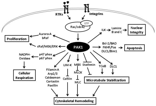
Importance of the field: P21-activated kinases (PAKs) are involved in multiple signal transduction pathways in mammalian cells. PAKs, and PAK1 in particular, play a role in such disorders as cancer, mental retardation and allergy. Cell motility, survival and proliferation, the organization and function of cytoskeleton and extracellular matrix, transcription and translation are among the processes affected by PAK1.

Areas covered in this review: We discuss the mechanisms that control PAK1 activity, its involvement in physiological and pathophysiological processes, the benefits and the drawbacks of the current tools to regulate PAK1 activity, the evidence that suggests PAK1 as a therapeutic target and the likely directions of future research.

What the reader will gain: The reader will gain a better knowledge and understanding of the areas described above.

Take home message: PAK1 is a promising therapeutic target in cancer and allergen-induced disorders. Its suitability as a target in vascular, neurological and infectious diseases remains ambiguous. Further advancement of this field requires progress on such issues as the development of specific and clinically acceptable inhibitors, the choice between targeting one or multiple PAK isoforms, elucidation of the individual roles of PAK1 targets and the mechanisms that may circumvent inhibition of PAK1.


Figures

Figure 1
Figure 1. Relative expression of PAK-1 mRNA in human tissue
Oligonucleotide microarray hybridization data has been obtained from www.biogps.com. Relative signal intensity values are depicted in arbitrary units with ranges.
Figure 2
Figure 2. Schematic representation of human group I and group II PAKs indicating the structural similarities and differences
Left upper panel show a 2.3 Ao resolution structure of human PAK1 (Group I) kinase domain from the available information on their crystal structures (retrieved from www.PyMOL.org). The asymmetric unit of the crystals contains two complexes, a auto-regulatory domain (aa 7-149) and a Kinase domain (aa 249-545) with a mutation in the kinase domain (K299R) that ablates its catalytic activity. These domains are linked into a dimer by the N-terminal segments (aa 78-88) of the auto-regulatory fragments. Right upper panel represents linear structure of Group I PAK isoforms. Group I PAKs contain a conserved overlapping PBD/CRIB/AID region. Interaction of Rac and cdc42 with the CRIB domain has been shown to release the group I PAKs from auto-inhibition. Left lower panel shows 1.6 Ao resolution structure of enzymatically active human PAK-4 catalytic domain (aa 291-591) when it is phosphorylated at the activating loop positions corresponding to Ser-474. It can be noted that the overlapping CRIB (aa 75-90) and AID (aa 83-149) regions that are present in group I PAKs is missing in PAK4. Right lower panel is a linear representation of group II PAKs. Group II PAKs lack AID, CRIB, acidic and PIX binding regions that are present in Group I PAKs.
Figure 3
Figure 3. Intracellular localization of PAK1
Fluorescent microscopic images of NIH 3T3 fibroblasts treated with control (PBS) and fibroblast growth factor (FGF; 20ng/ml) showing intracellular distribution of PAK1. In a resting cell, PAK1 is localized mostly in the cytoplasm. In order to study the PAK1 translocation in activated cells, NIH 3T3 fibroblasts were treated with 20ng/ml of FGF and incubated for 30 minutes in a CO2 incubator at 37°C. Cells were fixed with 2% para-formaldehyde and stained with PAK1 antibody (Santa Cruz biotechnology). Upon treatment with FGF, PAK1 is seen to be translocated to the plasma membrane according to its well-characterized role in the regulation of cytoskeletal dynamics and cell motility.
Figure 4
Figure 4. Schematic representation of PAK1 and some of its substrates regulating various cellular functions
Various stimuli, such as growth factors and integrins, activates PAK1 via GTP-bound Rac and Cdc42. Enhanced expression of Lamins B and C via phosphorylation and activation of Integrin Linked Kinase (ILK) has been implicated in the regulation of nuclear integrity. PAK1 is known to protect cells from apoptosis through at least three different pathways involving FKHR, Bcl-2 and DLC1. In the regulation of cellular proliferation, PAK1 regulates activity of Raf and Aurora kinases. PAK1 is also known to regulate cellular respiration via phoshorylations of p47- and p67- phox proteins. The most studied and well-characterized function of PAK1 is in the regulation of cytoskeletal remodeling. While PAK1 regulates microtubule dynamics through inhibition of stathmin and activation of TCoB and DLC1, it also regulates actin assembly and disassembly through phosphorylations of LIM Kinase and cytoskeletal proteins such as Filamin A, Arp2/3, Caldesmon, Cortactin and Paxillin. Apart from this, PAK1 also phosphorylates myosin binding unit (MBS) that in turn, inhibits phosphorylation of myosin light chain (MLC).

References

    1. Manser E, Leung T, Salihuddin H, Zhao ZS, Lim L. A brain serine/threonine protein kinase activated by Cdc42 and Rac1. Nature. 1994 Jan 6;367(6458):40–6. - PubMed
    1. Martin GA, Bollag G, McCormick F, Abo A. A novel serine kinase activated by rac1/CDC42Hs-dependent autophosphorylation is related to PAK65 and STE20. Embo J. 1995 May 1;14(9):1970–8. - PMC - PubMed
    1. Knaus UG, Morris S, Dong HJ, Chernoff J, Bokoch GM. Regulation of human leukocyte p21-activated kinases through G protein--coupled receptors. Science. 1995 Jul 14;269(5221):221–3. - PubMed
    1. Bagrodia S, Taylor SJ, Creasy CL, Chernoff J, Cerione RA. Identification of a mouse p21Cdc42/Rac activated kinase. J Biol Chem. 1995 Sep 29;270(39):22731–7. - PubMed
    1. Kumar A, Molli PR, Pakala SB, Nguyen TM Bui, Rayala SK, Kumar R. PAK thread from amoeba to mammals. Journal of cellular biochemistry. 2009 Jul 1;107(4):579–85. - PMC - PubMed

Publication types

MeSH terms