Review
. 2010 Jun 15;428(3):325-46.
doi: 10.1042/BJ20100046.

The cutting edge: membrane-anchored serine protease activities in the pericellular microenvironment

Affiliations
Review

The cutting edge: membrane-anchored serine protease activities in the pericellular microenvironment

Toni M Antalis et al. Biochem J. .

Abstract

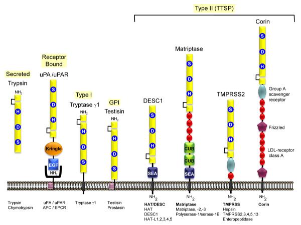
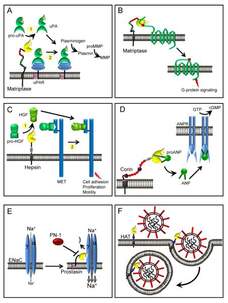
The serine proteases of the trypsin-like (S1) family play critical roles in many key biological processes including digestion, blood coagulation, and immunity. Members of this family contain N- or C-terminal domains that serve to tether the serine protease catalytic domain directly to the plasma membrane. These membrane-anchored serine proteases are proving to be key components of the cell machinery for activation of precursor molecules in the pericellular microenvironment, playing vital functions in the maintenance of homoeostasis. Substrates activated by membrane-anchored serine proteases include peptide hormones, growth and differentiation factors, receptors, enzymes, adhesion molecules and viral coat proteins. In addition, new insights into our understanding of the physiological functions of these proteases and their involvement in human pathology have come from animal models and patient studies. The present review discusses emerging evidence for the diversity of this fascinating group of membrane serine proteases as potent modifiers of the pericellular microenvironment through proteolytic processing of diverse substrates. We also discuss the functional consequences of the activities of these proteases on mammalian physiology and disease.


Figures

Figure 1
Figure 1. Membrane anchored serine proteases are linked directly to the plasma membrane
Most of the well characterized S1A serine proteases, such as the prototype enzymes trypsin and chymotrypsin, are secreted enzymes, associated with extracellular proteolysis. Some of these enzymes (uPA, urinary-type plasminogen activator; APC, activated protein C) may participate in pericellular proteolysis by binding to specific cell surface receptors (uPAR, uPA receptor; EPCR, endothelial protein C receptor). Tryptase γ1 is the only known type I transmembrane serine protease. The human GPI anchored serine proteases are prostasin and testisin. The type II transmembrane serine proteases (TTSP) are the largest group of pericellular serine proteases and may be divided into four subfamilies [12,21]: (i) the Human Airway Trypsin-like protease/Differentially Expressed in Squamous cell Carcinoma (HAT/DESC) subfamily for which the stem regions are all composed of a single SEA domain, (ii) the Hepsin/Transmembrane Protease, Serine (TMPRSS) subfamily, each of which have a group A scavenger receptor domain (SRCR) in their stem region, preceded by a single LDL receptor class A-like (LDLRA) domain in TMPRSS2, 3, 4, and 13 or in enteropeptidase, an array of SEA, LDLRA, CUB, and MAM domains, (iii) the Matriptase subfamily, each containing a SEA domain, two CUB domains, and 3–4 LDLRA domains in their stem region. Polyserase-1 is unique comprising two active, and one catalytically inactive serine protease domains and a stem region containing an LDLRA domain, and (iv) the Corin subfamily, consisting of a single member, corin, which possesses a complex stem region composed of two frizzled domains, eight LDLRA domains, and one SRCR domain.
Figure 2
Figure 2. Membrane anchored serine proteases participate in zymogen cascades
Pathways in which membrane anchored serine proteases have been shown to activate, or be activated by, serine proteases in vitro and in vivo. Proteases are color coded according to membrane localization sequences, (red) TTSPs, (green) GPI-anchored proteases, (blue) other secreted proteases. Lines indicate activation cleavages and loops indicate auto-activation. The dotted line indicates that hepsin is a weak activator of the matriptase zymogen. Membrane anchored serine proteases intersect the coagulation cascade (Factor VII activation), fibrinolysis (pro-uPA activation) and metalloproteinase pathways (pro-MMP-3 activation). Not shown is the activation of trypsinogen by enteropeptidase.
Figure 3
Figure 3. Proteolytic pathways associated with membrane anchored serine proteases in pericellular microenvironments
(A) Fibrinolysis. The membrane anchored serine proteases, matriptase, hepsin and Serase-1B may participate in the initiation of the plasminogen activation cascade via conversion of pro-uPA to active uPA. Matriptase is a relatively poor activator of pro-uPA in solution (1), whereas it is an effective activator of uPAR-bound pro-uPA and initiates pericellular plasminogen activation on the monocyte cell surface (2). (B) Inflammation. Matriptase and possibly other membrane serine proteases, may participate in the activation of PARs triggering G-protein signaling pathways. (C) Growth factor activation. Hepsin and matriptase may participate in the activation of pro-HGF to initiate signaling via the HGF receptor, MET, to modulate cell adhesion, proliferation and cell motility. Whether these enzymes target soluble pro-HGF (1) or receptor bound pro-HGF (2) is not known. (D) Natriuretic peptides. Corin processes pro-ANP to an active form that bind ANP receptors (ANPR) in the kidney and vasculature, increasing guanylate cyclase activity to regulate blood pressure and blood volume. (E) Sodium homeostasis. The open probability of the ENaC is increased by prostasin and TMPRSS4, resulting in increased cellular uptake of sodium (Na+) which in turn can regulate homeostasis of extracellular fluid volume, blood pressure and Na+ reabsorption. Processing of the α or γ subunits of ENaC is predicted to release an inhibitory fragment that leads to full activation of the channel. The serpin protease nexin-1 (PN-1, serpinE2) can inhibit prostasin mediated activation of ENaC. (F) Viral Pathogenicity. Several TTSPs, including HAT, TMPRSS2, 4, and 13 can process precursor viral proteins that facilitate virus entry and fusion into host cells.

References

    1. Ramsay AJ, Quesada V, Sanchez M, Garabaya C, Sarda MP, Baiget M, Remacha A, Velasco G, Lopez-Otin C. Matriptase-2 mutations in iron-refractory iron deficiency anemia patients provide new insights into protease activation mechanisms. Hum. Mol. Genet. 2009;18:3673–3683. - PubMed
    1. Neurath H, Walsh KA. Role of proteolytic enzymes in biological regulation (a review) Proc. Natl. Acad. Sci. U.S.A. 1976;73:3825–3832. - PMC - PubMed
    1. Hedstrom L. Serine protease mechanism and specificity. Chem. Rev. 2002;102:4501–4524. - PubMed
    1. Rawlings ND, Barrett AJ. Introduction: serine peptidases and their clans. In: Barrett AJ, Rawlings ND, Woessner JF, editors. In Handbook of Proteolytic Enzymes. 2nd Edition Vol. 2. Elsevier Ltd; London UK: 2004. pp. 1417–1439.
    1. Rawlings ND, Morton FR, Kok CY, Kong J, Barrett AJ. MEROPS: the peptidase database. Nucleic Acids Res. 2008;36:D320–D325. - PMC - PubMed

Publication types

Substances