Optic disc abnormalities - diagnosis, evolution and influence on visual acuity
- PMID: 20507293
- PMCID: PMC5509398
- DOI: 10.17305/bjbms.2010.2711
Optic disc abnormalities - diagnosis, evolution and influence on visual acuity
Abstract
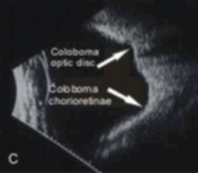
Congenital abnormalities of the optic disc are not so rare. The etiology for the most of them is unknown. Visual acuity of affected eye may be minimally or severely affected, depending on the extent of lesion. All of these conditions can be unilateral or bilateral. Children who have unilateral optic disc abnormalities generally present during the preschool years with sensory esotropia. Visual acuity may be unaffected like in optic disc pit, optic disc drusen, fibre medullares, ect. However, during the evolution they may cause a decrease in visual acuity like serous retinal detachment in optic disc pit, atrophy or subretinal neovascularisation in optic disc drusen. Some of them like fibre medullares needs only a good diagnose and they do not have any evolution. Fluorescein angiography and ultrasonography may be crucial diagnostic procedures to discover some of them, like optic disc drusen. Optic disc abnormalities may be associated with other congenital disorders of the eye and often central nervous system malformations. Secondary they may be associated retinal detachment, retinochisis, macular edema, choroid neovascularisation and lipid exudation. Some of these conditions may be found on routine ophthalmologic exam such as optic disc drusen and fibre medullares and often are diagnostically problem. The aim of our study was to present some of our cases with different optic disc abnormalities such as fibre medullares, optic disc coloboma, hypoplasio disc, optic disc drusen and optic disc pit.
Figures


References
-
- Azar N.F, Davis EA. Embryology of the eye. In: Yanoff M, Ducker JS, editors. Ophthalmology. second ed. Mosby; 2004. pp. 22–27.
-
- Pulifiato C, Hee M, Schuman JS, Fujimoto JG. Diseases of Optic nerve in: Optical chocherence tomography of ocular diseases. Elsever; 2007. pp. 372–373.
-
- Jandreck C. Optic nerve head anomalies. In: Heimann H, Kellner U, Foester HM, editors. Atlas of Fundus Angiography. Stuttgart. New York: Thieme; 2006. pp. 166–169.
-
- Brodsky MC. Congenital optic Disc Anomalities. In: Yanoff M, Ducker JS, editors. Ophthalmology. second ed. Mosby; 2004. pp. 1255–1258.
-
- Milenković S, Jaković N. Optic disc pit -Atipical optic disc coloboma. Medical Investigations. 2002;36(3):29–33.