

The constitutional t(11;22): implications for a novel mechanism responsible for gross chromosomal rearrangements
- PMID: 20507342
- PMCID: PMC3336963
- DOI: 10.1111/j.1399-0004.2010.01445.x
The constitutional t(11;22): implications for a novel mechanism responsible for gross chromosomal rearrangements
Abstract
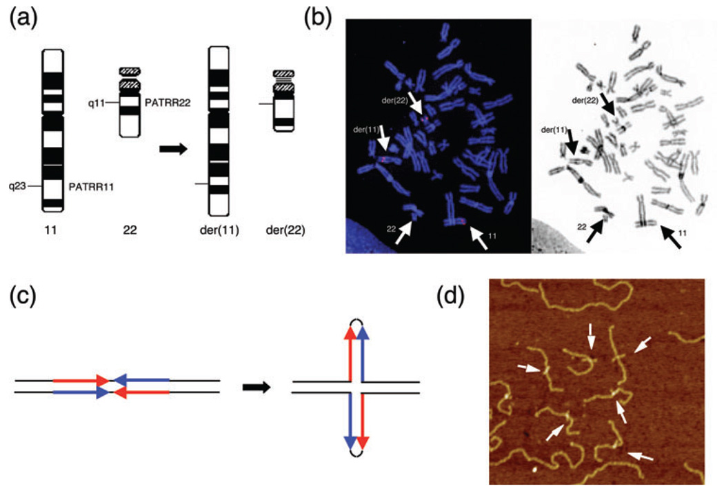
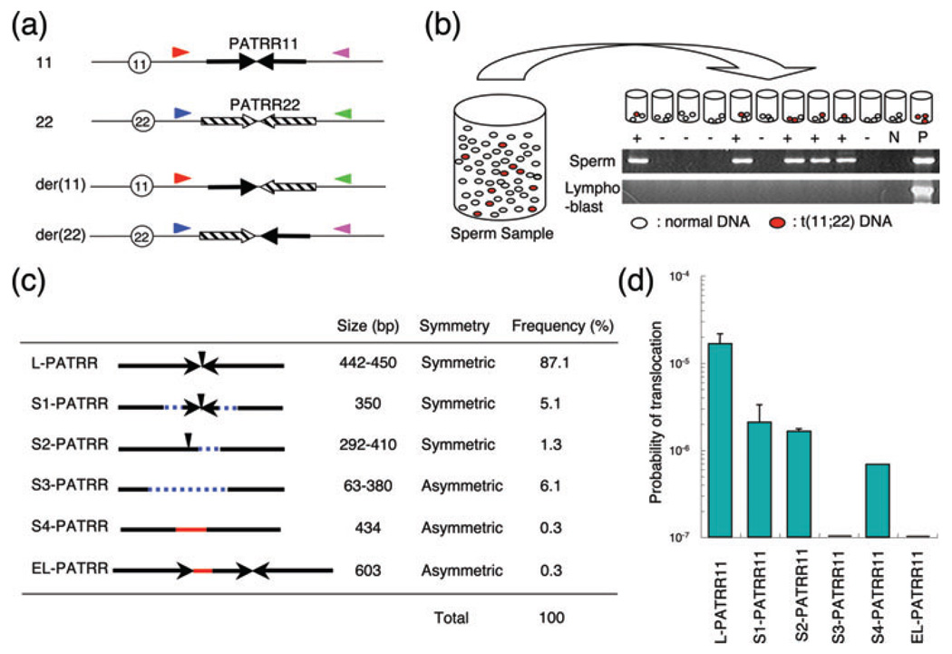
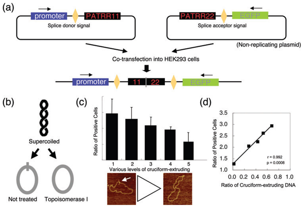
The constitutional t(11;22)(q23;q11) is the most common recurrent non-Robertsonian translocation in humans. The breakpoint sequences of both chromosomes are characterized by several hundred base pairs of palindromic AT-rich repeats (PATRRs). Similar PATRRs have also been identified at the breakpoints of other nonrecurrent translocations, suggesting that PATRR-mediated chromosomal translocation represents one of the universal pathways for gross chromosomal rearrangement in the human genome. We propose that PATRRs have the potential to form cruciform structures through intrastrand-base pairing in single-stranded DNA, creating a source of genomic instability and leading to translocations. Indeed, de novo examples of the t(11;22) are detected at a high frequency in sperm from normal healthy males. This review synthesizes recent data illustrating a novel paradigm for an apparent spermatogenesis-specific translocation mechanism. This observation has important implications pertaining to the predominantly paternal origin of de novo gross chromosomal rearrangements in humans.
© 2010 John Wiley & Sons A/S.
Conflict of interest statement
We declare no conflict of interest.
Figures
Similar articles
-
Chromosomal translocations and palindromic AT-rich repeats.Curr Opin Genet Dev. 2012 Jun;22(3):221-8. doi: 10.1016/j.gde.2012.02.004. Epub 2012 Mar 6. Curr Opin Genet Dev. 2012. PMID: 22402448 Free PMC article. Review.
-
Palindrome-mediated chromosomal translocations in humans.DNA Repair (Amst). 2006 Sep 8;5(9-10):1136-45. doi: 10.1016/j.dnarep.2006.05.035. Epub 2006 Jul 10. DNA Repair (Amst). 2006. PMID: 16829213 Free PMC article. Review.
-
Long AT-rich palindromes and the constitutional t(11;22) breakpoint.Hum Mol Genet. 2001 Nov 1;10(23):2605-17. doi: 10.1093/hmg/10.23.2605. Hum Mol Genet. 2001. PMID: 11726547
-
Paternal origin of the de novo constitutional t(11;22)(q23;q11).Eur J Hum Genet. 2010 Jul;18(7):783-7. doi: 10.1038/ejhg.2010.20. Epub 2010 Feb 24. Eur J Hum Genet. 2010. PMID: 20179746 Free PMC article.
-
Cruciform extrusion propensity of human translocation-mediating palindromic AT-rich repeats.Nucleic Acids Res. 2007;35(4):1198-208. doi: 10.1093/nar/gkm036. Epub 2007 Jan 30. Nucleic Acids Res. 2007. PMID: 17264116 Free PMC article.
Cited by
-
Supernumerary derivative 22 chromosome resulting from novel constitutional non-Robertsonian translocation: t(20;22)-Case Report.Mol Cytogenet. 2022 Mar 26;15(1):14. doi: 10.1186/s13039-022-00591-4. Mol Cytogenet. 2022. PMID: 35346304 Free PMC article.
-
Breakpoint analysis of the recurrent constitutional t(8;22)(q24.13;q11.21) translocation.Mol Cytogenet. 2014 Aug 13;7:55. doi: 10.1186/s13039-014-0055-x. eCollection 2014. Mol Cytogenet. 2014. PMID: 25478009 Free PMC article.
-
The cellular etiology of chromosome translocations.Curr Opin Cell Biol. 2013 Jun;25(3):357-64. doi: 10.1016/j.ceb.2013.02.015. Epub 2013 Mar 14. Curr Opin Cell Biol. 2013. PMID: 23498663 Free PMC article. Review.
-
Non-B DB v2.0: a database of predicted non-B DNA-forming motifs and its associated tools.Nucleic Acids Res. 2013 Jan;41(Database issue):D94-D100. doi: 10.1093/nar/gks955. Epub 2012 Nov 3. Nucleic Acids Res. 2013. PMID: 23125372 Free PMC article.
-
Molecular cytogenetic characterization of partial trisomy of the long arm of chromosome 11 in a patient with multiple congenital anomalies.Mol Cytogenet. 2022 Apr 19;15(1):17. doi: 10.1186/s13039-022-00595-0. Mol Cytogenet. 2022. PMID: 35440058 Free PMC article. Review.
References
-
- Zackai EH, Emanuel BS. Site-specific reciprocal translocation, t(11;22) (q23;q11), in several unrelated families with 3:1 meiotic disjunction. Am J Med Genet. 1980;7:507–521. - PubMed
-
- Fraccaro M, Lindsten J, Ford CE, et al. The 11q;22q translocation: a European collaborative analysis of 43 cases. Hum Genet. 1980;56:21–51. - PubMed
-
- Kurahashi H, Bolor H, Kato T, et al. Recent advance in our understanding of the molecular nature of chromosomal abnormalities. J Hum Genet. 2009;54:253–260. - PubMed
Publication types
MeSH terms
Substances
Grants and funding
LinkOut - more resources
Full Text Sources
