

Minocycline inhibits glial proliferation in the H-Tx rat model of congenital hydrocephalus
- PMID: 20507614
- PMCID: PMC2889858
- DOI: 10.1186/1743-8454-7-7
Minocycline inhibits glial proliferation in the H-Tx rat model of congenital hydrocephalus
Abstract
Background: Reactive astrocytosis and microgliosis are important features of the pathophysiology of hydrocephalus, and persistent glial "scars" that form could exacerbate neuroinflammation, impair cerebral perfusion, impede neuronal regeneration, and alter biomechanical properties. The purpose of this study was to determine the efficacy of minocycline, an antibiotic known for its anti-inflammatory properties, to reduce gliosis in the H-Tx rat model of congenital hydrocephalus.
Methods: Minocycline (45 mg/kg/day i.p. in 5% sucrose at a concentration of 5-10 mg/ml) was administered to hydrocephalic H-Tx rats from postnatal day 15 to day 21, when ventriculomegaly had reached moderate to severe stages. Treated animals were compared to age-matched non-hydrocephalic and untreated hydrocephalic littermates. The cerebral cortex (both gray matter laminae and white matter) was processed for immunohistochemistry (glial fibrillary acidic protein, GFAP, for astrocytes and ionized calcium binding adaptor molecule, Iba-1, for microglia) and analyzed by qualitative and quantitative light microscopy.
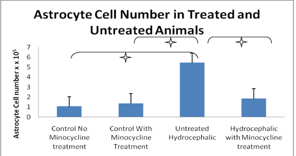
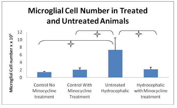
Results: The mean number of GFAP-immunoreactive astrocytes was significantly higher in untreated hydrocephalic animals compared to both types of controls (p < 0.001). Minocycline treatment of hydrocephalic animals reduced the number of GFAP immunoreactive cells significantly (p < 0.001). Likewise, the mean number of Iba-1 immunoreactive microglia was significantly higher in untreated hydrocephalic animals compared to both types of controls (p < 0.001). Furthermore, no differences in the numbers of GFAP-positive astrocytes or Iba-1-positive microglia were noted between control animals receiving no minocycline and control animals receiving minocycline, suggesting that minocycline does not produce an effect under non-injury conditions. Additionally, in six out of nine regions sampled, hydrocephalic animals that received minocycline injections had significantly thicker cortices when compared to their untreated hydrocephalic littermates.
Conclusions: Overall, these data suggest that minocycline treatment is effective in reducing the gliosis that accompanies hydrocephalus, and thus may provide an added benefit when used as a supplement to ventricular shunting.
Figures
Similar articles
-
Minocycline reduces reactive gliosis in the rat model of hydrocephalus.BMC Neurosci. 2012 Dec 5;13:148. doi: 10.1186/1471-2202-13-148. BMC Neurosci. 2012. PMID: 23217034 Free PMC article.
-
Reduction of astrogliosis and microgliosis by cerebrospinal fluid shunting in experimental hydrocephalus.Cerebrospinal Fluid Res. 2007 Jun 7;4:5. doi: 10.1186/1743-8454-4-5. Cerebrospinal Fluid Res. 2007. PMID: 17555588 Free PMC article.
-
Reduced subventricular zone proliferation and white matter damage in juvenile ferrets with kaolin-induced hydrocephalus.Exp Neurol. 2013 Oct;248:112-28. doi: 10.1016/j.expneurol.2013.06.004. Epub 2013 Jun 12. Exp Neurol. 2013. PMID: 23769908
-
Reactive astrocytosis, microgliosis and inflammation in rats with neonatal hydrocephalus.Exp Neurol. 2010 Nov;226(1):110-9. doi: 10.1016/j.expneurol.2010.08.010. Epub 2010 Aug 14. Exp Neurol. 2010. PMID: 20713048
-
Reactive gliosis and neuroinflammation in rats with communicating hydrocephalus.Neuroscience. 2012 Aug 30;218:317-25. doi: 10.1016/j.neuroscience.2012.05.004. Epub 2012 May 11. Neuroscience. 2012. PMID: 22583796
Cited by
-
Inhibition of Wnt/β-catenin signal is alleviated reactive gliosis in rats with hydrocephalus.Childs Nerv Syst. 2015 Feb;31(2):227-34. doi: 10.1007/s00381-014-2613-2. Epub 2015 Jan 7. Childs Nerv Syst. 2015. PMID: 25564198
-
Iron-Induced Hydrocephalus: the Role of Choroid Plexus Stromal Macrophages.Transl Stroke Res. 2023 Apr;14(2):238-249. doi: 10.1007/s12975-022-01031-6. Epub 2022 May 11. Transl Stroke Res. 2023. PMID: 35543803 Free PMC article.
-
Effects of minocycline on epiplexus macrophage activation, choroid plexus injury and hydrocephalus development in spontaneous hypertensive rats.J Cereb Blood Flow Metab. 2019 Oct;39(10):1936-1948. doi: 10.1177/0271678X19836117. Epub 2019 Mar 12. J Cereb Blood Flow Metab. 2019. PMID: 30862302 Free PMC article.
-
Minocycline reduces reactive gliosis in the rat model of hydrocephalus.BMC Neurosci. 2012 Dec 5;13:148. doi: 10.1186/1471-2202-13-148. BMC Neurosci. 2012. PMID: 23217034 Free PMC article.
-
Nonsurgical therapy for hydrocephalus: a comprehensive and critical review.Fluids Barriers CNS. 2016 Feb 5;13:3. doi: 10.1186/s12987-016-0025-2. Fluids Barriers CNS. 2016. PMID: 26846184 Free PMC article. Review.
References
LinkOut - more resources
Full Text Sources
Miscellaneous
