. 2010 Sep;20(9):1170-85.
doi: 10.1093/glycob/cwq083. Epub 2010 May 27.

Human L-selectin preferentially binds synthetic glycosulfopeptides modeled after endoglycan and containing tyrosine sulfate residues and sialyl Lewis x in core 2 O-glycans

Affiliations

Human L-selectin preferentially binds synthetic glycosulfopeptides modeled after endoglycan and containing tyrosine sulfate residues and sialyl Lewis x in core 2 O-glycans

Anne Leppänen et al. Glycobiology. 2010 Sep.

Abstract

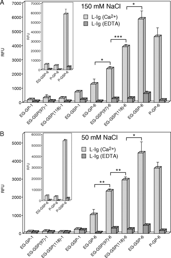
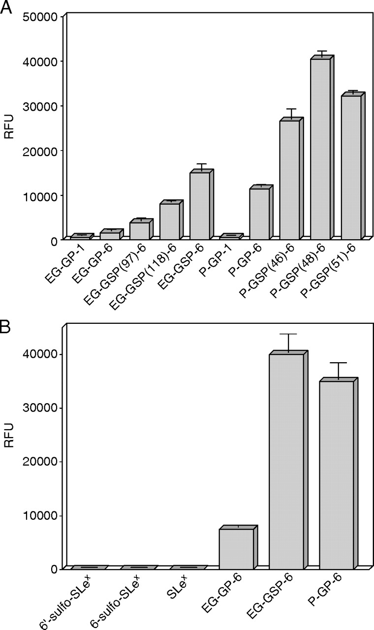
Endoglycan is a mucin-like glycoprotein expressed by endothelial cells and some leukocytes and is recognized by L-selectin, a C-type lectin important in leukocyte trafficking and extravasation during inflammation. Here, we show that recombinant L-selectin and human T lymphocytes expressing L-selectin bind to synthetic glycosulfopeptides (GSPs). These synthetic glycosulfopeptides contain 37 amino acid residues modeled after the N-terminus of human endoglycan and contain one or two tyrosine sulfates (TyrSO(3)) along with a nearby core-2-based Thr-linked O-glycan with sialyl Lewis x (C2-SLe(x)). TyrSO(3) at position Y118 was more critical for binding than at Y97. C2-SLe(x) at T124 was required for L-selectin recognition. Interestingly, under similar conditions, neither L-selectin nor T lymphocytes showed appreciable binding to the sulfated carbohydrate epitope 6-sulfo-SLe(x). P-selectin also bound to endoglycan-based GSPs but with lower affinity than toward GSPs modeled after PSGL-1, the physiological ligand for P- and L-selectin that is expressed on leukocytes. These results demonstrate that TyrSO(3) residues in association with a C2-SLe(x) moiety within endoglycan and PSGL-1 are preferentially recognized by L-selectin.


Figures

Fig. 1
Fig. 1
The structures of glyco(sulfo)peptides and sugars used in the present study. Tyrosine sulfate and O-glycan attachment sites are numbered according to the sequence of human endoglycan (EG) or PSGL-1 (P). The number in the parenthesis indicates the position of tyrosine sulfate residues, and the last number indicates the number of sugar residues. All peptides contain an extra C-terminal cysteine residue
Fig. 2
Fig. 2
Binding of L-sel-Ig to immobilized glyco(sulfo)peptides in a fluorescence-based solid-phase assay at physiologic and low salt buffer. Biotinylated glyco(sulfo)peptides were immobilized on streptavidin-coated microtiter wells (A) 20 pmol/well; (B) 10 pmol/well). (A) L-sel-Ig (40 μg/ml) was incubated with the immobilized GSPs in physiologic salt buffer (20 mM MOPS, pH 7.5, 150 mM NaCl, 1% BSA, 0.05% Tween 20, 0.02% NaN3) containing 2 mM CaCl2 and 2 mM MgCl2 (light gray bars) or 5 mM EDTA (dark gray bars). (B) L-sel-Ig (20 μg/ml) was incubated with the immobilized GSPs in low salt buffer (20 mM MOPS, pH 7.5, 50 mM NaCl, 1% BSA, 0.05% Tween 20, 0.02% NaN3) containing 2 mM CaCl2 and 2 mM MgCl2 (light gray bars) or 5 mM EDTA (dark gray bars). Fluorescently labeled anti-human IgG was used to detect the bound L-sel-Ig. The inset shows binding of L-sel-Ig to immobilized P-GSP-6. All assays were performed in triplicate, and the results represent the mean ± SD of three determinations. *P < 0.05; **P < 0.01; ***P < 0.001
Fig. 3
Fig. 3
Binding of L-sel-Ig to immobilized glyco(sulfo)peptides and sugars in a fluorescence-based solid-phase assay at low salt buffer. Biotinylated glyco(sulfo)peptides and sugars were immobilized on streptavidin-coated microtiter wells (10 pmol/well). L-sel-Ig (A) 20 μg/ml or (B) 40 μg/ml was incubated with the immobilized ligands in low salt buffer (20 mM MOPS, pH 7.5, 50 mM NaCl, 2 mM CaCl2, 2 mM MgCl2, 1% BSA, 0.05% Tween 20, 0.02% NaN3). Fluorescently labeled anti-human IgG was used to detect the bound L-sel-Ig. All assays were performed in triplicate, and the results represent the mean ± SD of three determinations
Fig. 4
Fig. 4
Inhibition of L-sel-Ig binding to immobilized glyco(sulfo)peptides by anti-EG peptide antibodies. Biotinylated glyco(sulfo)peptides were immobilized on streptavidin-coated microtiter wells (5 pmol/well). Anti-QPP and anti-SGF antibodies (100 μg/ml) were first incubated with the immobilized GSPs in PBS containing 1% BSA and 0.05% Tween 20, and unbound antibodies were removed by washing. L-sel-Ig (10 μg/ml) was preincubated with fluorescently labeled anti-human IgG in low salt buffer (20 mM MOPS, pH 7.5, 50 mM NaCl, 2 mM CaCl2, 2 mM MgCl2, 1% BSA, 0.05% Tween 20, 0.02% NaN3) before adding to the wells. Assays with anti-QPP and without an antibody were performed in triplicate, assays with anti-SGF were performed in duplicate, and the results represent the mean ± SD of three or two determinations, respectively
Fig. 5
Fig. 5
Affinity chromatography of glyco(sulfo)peptides on immobilized L-sel-Ig at low salt buffer. The indicated radiolabeled glyco(sulfo)peptides were loaded into the L-sel-Ig column in low salt buffer (20 mM MOPS, 50 mM NaCl, 0.02% NaN3, pH 7.5) containing 2 mM CaCl2 and 2 mM MgCl2 (A), (B) and (D) or 1 mM EDTA (C). In (A), dashed lines represent control experiments with the indicated glycosulfopeptides where chromatography was performed in buffer containing 1 mM EDTA. The arrow indicates the fraction number (21) where 10 mM EDTA was used to replace divalent cations in buffer. Experiments shown are representative of three independent experiments
Fig. 6
Fig. 6
Equilibrium-binding affinity of L-sel-Ig for P-GSP-6, P-GP-6 and EG-GSP-6 in a fluorescence-based solid-phase assay at low salt buffer. Biotinylated glyco(sulfo)peptides were immobilized on streptavidin-coated microtiter wells (10 pmol/well). Various concentrations of L-sel-Ig were incubated with the immobilized (A) P-GSP-6, (B) P-GP-6 and (C) EG-GSP-6 in low salt buffer (20 mM MOPS, pH 7.5, 50 mM NaCl, 2 mM CaCl2, 2 mM MgCl2, 1% BSA, 0.05% Tween 20, 0.02% NaN3). Fluorescently labeled anti-human IgG (40 μg/ml) was used to detect the bound L-sel-Ig. Assays were performed in triplicate (A) and (B) or in duplicate (C), and the results represent the mean ± standard error of the mean. Experiments shown are representative of three independent experiments
Fig. 7
Fig. 7
Binding of human T lymphocytes to immobilized glyco(sulfo)peptides and sugars in a fluorescence-based solid-phase assay at physiologic buffer. Biotinylated glyco(sulfo)peptides and sugars were immobilized on streptavidin-coated microtiter wells (10 pmol/well). Purified, fluorescently labeled human T lymphocytes (A) 190,000 cells/well or (B) 65,000 cells/well were incubated with the immobilized ligands in Hank's balanced salt solution (with Ca2 and Mg2) containing 1% BSA (light gray bars). (B) In control experiments, a function-blocking mAb to L-selectin, DREG-56 (20 μg/ml) (medium gray bars), or 5 mM EDTA (dark gray bars) were preincubated with the cells in HBSS before adding to the wells. The inset in (A) shows binding of T lymphocytes to immobilized P-GSP-6. All assays were performed in triplicate, and the results represent the mean ± SD of three determinations. Experiment shown in (A) is a representative of eight independent experiments using T cells isolated from five different donors. *P < 0.05; **P < 0.01
Fig. 8
Fig. 8
Binding of P-sel-Ig to immobilized sugars and glyco(sulfo)peptides in a fluorescence-based solid-phase assay at physiologic buffer. Biotinylated sugars and glyco(sulfo)peptides were immobilized on streptavidin-coated microtiter wells (10 pmol/well). P-sel-Ig (20 μg/ml) was incubated with the immobilized GSPs in physiologic salt buffer (20 mM MOPS, pH 7.5, 150 mM NaCl, 1% BSA, 0.05% Tween 20, 0.02% NaN3) containing 2 mM CaCl2 and 2 mM MgCl2 (light gray bars) or 5 mM EDTA (dark gray bars). Fluorescently labeled anti-human IgG was used to detect the bound P-sel-Ig. All assays were performed in triplicate, and the results represent the mean ± SD of three determinations. Experiment shown is a representative of three independent experiments. *P < 0.05; **P < 0.01

References

    1. Baumhueter S, Singer MS, Henzel W, Hemmerich S, Renz M, Rosen SD, Lasky LA. Binding of L-selectin to the vascular sialomucin CD34. Science. 1993;262:436–438. - PubMed
    1. Berg EL, McEvoy LM, Berlin C, Bargatze RF, Butcher EC. L-selectin-mediated lymphocyte rolling on MAdCAM-1. Nature. 1993;366:695–698. - PubMed
    1. Berg EL, Robinson MK, Warnock RA, Butcher EC. The human peripheral lymph node vascular addressin is a ligand for LECAM-1, the peripheral lymph node homing receptor. J Cell Biol. 1991;114:343–349. - PMC - PubMed
    1. Bernimoulin MP, Zeng XL, Abbal C, Giraud S, Martinez M, Michielin O, Schapira M, Spertini O. Molecular basis of leukocyte rolling on PSGL-1. Predominant role of core-2 O-glycans and of tyrosine sulfate residue 51. J Biol Chem. 2003;278:37–47. - PubMed
    1. Bistrup A, Bhakta S, Lee JK, Belov YY, Gunn MD, Zuo FR, Huang CC, Kannagi R, Rosen SD, Hemmerich S. Sulfotransferases of two specificities function in the reconstitution of high endothelial cell ligands for L-selectin. J Cell Biol. 1999;145:899–910. - PMC - PubMed

Publication types

MeSH terms